 | Info
Sheets |
| | | | | | | | | | | | | | | | | | | | | | | | |
 | Out-
side |
| | | | |
|
| | | | |
Result : Searchterm 'Tip Angle' found in 3 terms [ ] and 2 definitions [ ] and 2 definitions [ ], (+ 11 Boolean[ ], (+ 11 Boolean[ ] results ] results
| | previous 6 - 10 (of 16) nextResult Pages : [1] [2 3 4] |  | |  | Searchterm 'Tip Angle' was also found in the following service: | | | | |
| |  |
| |
|
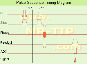
In simple ultrafast GRE imaging, TR and TE are so short, that tissues have a poor imaging signal and - more importantly - poor contrast except when contrast media enhanced ( contrast enhanced angiography). Therefore, the magnetization is 'prepared' during the preparation module, most frequently by an initial 180° inversion pulse.
In the pulse sequence timing diagram, the basic ultrafast gradient echo sequence is illustrated. The 180° inversion pulse is executed one time (to the left of the vertical line), the right side represents the data collection period and is often repeated depending on the acquisition parameters.
See also Pulse Sequence Timing Diagram, there you will find a description of the components.
Ultrafast GRE sequences have a short TR,TE, a low flip angle and TR is so short that image acquisition lasts less than 1 second and typically less than 500 ms. Common TR: 3-5 msec, TE: 2 msec, and the flip angle is about 5°.
Such sequences are often labeled with the prefix 'Turbo' like TurboFLASH, TurboFFE and TurboGRASS.
This allows one to center the subsequent ultrafast GRE data acquisition around the inversion time TI, where one of the tissues of interest has very little signal as its z-magnetization is passing through zero.
Unlike a standard inversion recovery (IR) sequence, all lines or a substantial segment of k-space image lines are acquired after a single inversion pulse, which can then together be considered as readout module. The readout module may use a variable flip angle approach, or the data acquisition may be divided into mul tiple segments (shots). The latter is useful particularly in cardiac imaging where acquiring all lines in a single segment may take too long relative to the cardiac cycle to provide adequate temporal resolution.
If mul tiple lines are acquired after a single pulse, the pulse sequence is a type of gradient echo echo planar imaging (EPI) pulse sequence. See also Magnetization Prepared Rapid Gradient Echo ( MPRAGE) and Turbo Field Echo ( TFE). | | | | | | | | | | |
| | | | | |
| |
|
(SAR) The Specific Absorption Rate is defined as the RF power absorbed per unit of mass of an object, and is measured in watts per kilogram (W/kg).
The SAR describes the potential for heating of the patient's tissue due to the application of the RF energy necessary to produce the MR signal. Inhomogeneity of the RF field leads to a local exposure where most of the absorbed energy is applied to one body region rather than the entire person, leading to the concept of a local SAR. Hot spots may occur in the exposed tissue, to avoid or at least minimize effects of such theoretical complications, the frequency and the power of the radio frequency irradiation should be kept at the lowest possible level. Averaging over the whole body leads to the global SAR.
It increases with field strength, radio frequency power and duty cycle, transmitter-coil type and body size. The doubling of the field strength from 1.5 Tesla (1.5T) to 3 Tesla ( 3T) leads to a quadrupling of SAR. In high and ultrahigh fields, some of the mul tiple echo, mul tiple-slice pulse sequences may create a higher SAR than recommended by the agencies. SAR can be reduced by lower flip angle and longer repetition times, which could potentially affect image contrast.
Normally no threatening increase in temperature could be shown. Even in high magnetic fields, the local temperature increases not more than 1°C. 2.1°C is the highest measured increase in skin temperature. Eddy currents may heat up implants and thus may cause local heating.
FDA SAR limits:
•
Whole body: 4W/kg/15-minute exposure averaged;
•
Head: 3W/kg/10-minute exposure averaged;
•
Head or torso: 8W/kg/5 minute exposure per gram of tissue;
•
Extremities: 12W/kg/5 minute exposure per gram of tissue.
IEC (International Electrotechnical Commission) SAR limits of some European countries:
All limits are averaged over 6 minutes.
•
Level 0 (normal operating mode): Whole body 2W/kg; Head 3.2W/kg; Head or Torso (local) 10W/kg;
Extremities (local) 20W/kg;
•
Level I (first level controlled operating mode): Whole body 4W/kg; Head 3.2W/kg; Head or Torso (local) 10W/kg; Extremities (local) 20W/kg;
•
Level II (second level controlled operating mode): All values are over Level I values.
(For more details: IEC 60601-2-33 (2002))
In most countries standard MRI systems are limited to a maximum SAR of 4 W/kg, so most scanning in level II is impossible.
For Level I, in addition to routine monitoring, particular caution must be exercised for patients who are sensitive to temperature increases or to RF energy.
For Japan different SAR limits are valid. | | | |
• View the DATABASE results for 'Specific Absorption Rate' (8).
| | |
• View the NEWS results for 'Specific Absorption Rate' (1).
| | | | |  Further Reading: Further Reading: | | Basics:
|
|
News & More:
| |
| |
| | | | | |
| |
|
A coil of wire wound in the form of a long cylinder. When a current is passed through the coil, the magnetic field within the coil is relatively uniform. Solenoid RF coils are commonly used when the static magnetic field is perpendicular to the long axis of the body.
The reason against a solenoidal coil is that the signal is missed when the axis of the solenoidal coil is coaxial with the horizontal magnetic field Bo. If the coil's axis can be put near a right angle (60° to 90°) with the principal field, so the loss in the signal is greatly dismissed, and good SNR can be achieved.
The two forms are single turn solenoid and mul tiple turn solenoids. | | | | | |
| | | Searchterm 'Tip Angle' was also found in the following service: | | | | |
| | |
| |
|
Contrast enhanced GRE sequences provide T2 contrast but have a relatively poor SNR. Repetitive RF pulses with small flip angles together with appropriate gradient profiles lead to the superposition of two resonance signals.
The first signal is due to the free induction decay FID observed after the first and all ensuing RF excitations.
The second is a resonance signal obtained as a result of a spin echo generated by the second and all addicted RF-pulses.
Hence it is absent after the first excitation, it is a result of the free induction decay of the second to last RF-excitation and has a TE, which is almost 2TR.
For this echo to occur the gradients have to be completely symmetrical relative to the half time between two RF-pulses, a condition that makes it difficult to integrate this pulse sequence into a multiple slice imaging technique.
The second signal not only contains echo contributions from free induction decay, but obviously weakened by T2-decay.
Since the echo is generated by a RF-pulse, it is truly T2 rather than T2* weighted. Correspondingly it is also less sensitive to susceptibility changes and field inhomogeneities.
Companies use different acronyms to describe certain techniques.
Different terms (see also acronyms) for these gradient echo pulse sequences:
CE-FAST Contrast Enhanced Fourier Acquired Steady State,
CE-FFE Contrast Enhanced Fast Field Echo,
CE-GRE Contrast Enhanced Gradient-Echo,
DE-FGR Driven Equilibrium FGR,
FADE FASE Acquisition Double Echo,
PSIF Reverse Fast Imaging with Steady State Precession,
SSFP Steady State Free Precession,
T2 FFE Contrast Enhanced Fast Field Echo (T2 weighted).
In this context, 'contrast enhanced' refers to the pulse sequence, it does not mean enhancement with a contrast agent. | | | |
• View the DATABASE results for 'Contrast Enhanced Gradient Echo Sequence' (4).
| | | | |
| | | | | |
| |
|
If available, some graphic aids can be helpful to show image orientations.
1) A graphic icon of the labeled primary axes (A, L, H) with relative lengths given by direction sines and orientation as if viewed from the normal to the image plane can help orient the viewer, both to identify image plane orientation and to indicate possible in plane rotation.
2) Ingraphic prescription of obliques from other images, a sample original image with an overlaid line or set of lines indicating the intersection of the original and oblique image planes can help orient the viewer.
•
The basic anatomical directions are:
right(R) to left (L), posterior (P) to anterior (A), and feet (F) to head (H).
•
A standard display orientation for images in the basic slice orientation is:
1) transverse: A to top of image and L to right,
2) coronal: H to top of image and L to right and
3) sagittal: H to top of image and A to left.
The location in the R/L and P/A directions can be specified relative to the axis of the magnet.
The F/H location can be specified relative to a convenient patient structure.
The orientation of single oblique slices can be specified by rotating a slice in one of the basic orientations toward one of the other two basic orthogonal planes about an axis defined by the intersection of the 2 planes.
Double oblique slices can be specified as the result of tipping a single oblique plane toward the remaining basic orientation plane, about an axis defined by the intersection of the oblique plane and the remaining basic plane. In double oblique angulations, the first rotation is chosen about the vertical image axis and the second about the (new) horizontal axis.
Angles are chosen to have magnitudes less than 90° (for single oblique slices less than 45°); the sign of the angle is taken to be positive when the rotation brings positive axes closer together. | | | | | |
• View the DATABASE results for 'Orientation' (16).
| | | | |
| | | | |
| | |
| | | |
|
| |
 | Look
Ups |
| |


