 | Info
Sheets |
| | | | | | | | | | | | | | | | | | | | | | | | |
 | Out-
side |
| | | | |
|
| | | | | |  | Searchterm 'Range' was also found in the following services: | | | | |
|  |  |
| |
|
Often used to indicate an image where most of the contrast between tissues or tissue states is due to differences in tissue T2 created typically by using longer TE and TR times.
This term may be misleading in that the potentially important effects of tissue density differences and the range of tissue T2 values are often ignored.
Choosing the machine parameters such that TR greater than T1 (typically greater than 2 000 ms) and TE less than T2 (typically greater than 100 ms) and noting that (1-exp(-TR/T1) = 1 for TR/T1 much greater than 1, will reduce Eq. 1 to the expression
Mxy = Mxy0exp(-TE/T2)
which is dependent on T2 only, hence the term T2 weighting.
Therefore T2 weighted image contrast state is approached by imaging with a TR long compared to tissue T1 (to reduce T1 contribution to image contrast) and a TE between the longest and shortest tissue T2s of interest. A TR greater than 3 times the longest T1 is required for the T1 effect to be less than 5%. Due to the wide range of T1 and T2 and tissue density values that can be found in the body, an image that is T2 weighted for some tissues may not be so for others.
See also T2 Time.
Lesions with short T2 are (dark in T2 weighted sequences): acute haemorrhage (deoxyHb)
haemosiderin
physiologic iron (basal ganglia, etc.)
mucinous lesions. | | | |  | | | | | | | | |  Further Reading: Further Reading: | | Basics:
|
|
News & More:
| |
| |
|  | |  |  |  |
| |
|
(3D MRA) The 3D angiography technique can be applied to focus on fast flowing (arterial) blood and to visualize small tortuous vessels. 3D TOF images are less sensitive to turbulent flow artifacts.
The advantage of this approach is that the signal, acquired from the entire
volume has an increased signal to noise ratio. Slices are defined by a second phase encoded axis, which divides the volume into 'partitions'.
3D TOF MRA is acquired with 3D FT slabs or multiple overlapping thin 3D FT slabs ( MOTSA) depending on the coverage required and the range of flow-velocities under examination.
Such 3D techniques can provide equal spatial resolution along all three axes, i.e. be 'isotropic', or the partition thickness can be greater or less than the in plane spatial resolution in which case can be said to be 'anisotropic'.
The circle of Willis, anatomy as well as its fast arterial flow, lends itself well to both 3D TOF and 2D or 3D phase contrast angiography. | | | |  | |
• View the DATABASE results for '3 Dimensional Magnetic Resonance Angiography' (2).
| | | | |  Further Reading: Further Reading: | Basics:
|
|
| |
|  | |  |  |  |
| |
|

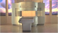
From Hitachi Medical Systems America Inc.;
the AIRIS II, an entry in the diagnostic category of open MR systems, was designed by Hitachi
Medical Systems America Inc. (Twinsburg, OH, USA) and Hitachi Medical Corp. (Tokyo) and is manufactured by the Tokyo branch. A 0.3 T field-strength magnet and phased array coils deliver high image quality without the need for a tunnel-type high-field system, thereby significantly improving patient comfort not only for claustrophobic patients.
Device Information and Specification
CLINICAL APPLICATION
Whole body
QD Head, MA Head and Neck, QD C-Spine, MA or QD Shoulder, MA CTL Spine, QD Knee, Neck, QD TMJ, QD Breast, QD Flex Body (4 sizes), Small and Large Extrem., QD Wrist, MA Foot and Ankle (WIP), PVA (WIP)
SE, GE, GR, IR, FIR, STIR, FSE, ss-FSE, FLAIR, EPI -DWI, SE-EPI, ms - EPI, SSP, MTC, SARGE, RSSG, TRSG, MRCP, Angiography: CE, 2D/3D TOF
IMAGING MODES
Single, multislice, volume study
TR
SE: 30 - 10,000msec GE: 20 - 10,000msec IR: 50 - 16,700msec FSE: 200 - 16,7000msec
TE
SE : 10 - 250msec IR: 10 -250msec GE: 5 - 50 msec FSE: 15 - 2,000
0.05 sec/image (256 x 256)
2D: 2 - 100 mm; 3D: 0.5 - 5 mm
Level Range: -2,000 to +4,000
POWER REQUIREMENTS
208/220/240 V, single phase
COOLING SYSTEM TYPE
Air-cooled
2.0 m lateral, 2.5 m vert./long
| |  | |
• View the DATABASE results for 'AIRIS II™' (2).
| | | | |
|  |  | Searchterm 'Range' was also found in the following services: | | | | |
|  |  |
| |
|

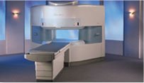
From Hitachi Medical Systems America, Inc.;
the AIRIS made its debut in 1995. Hitachi followed up with the AIRIS II system, which has proven equally successfully. 'All told, Hitachi has installed more than 1,000 MRI systems in the U.S., holding more than 17 percent of the total U.S. MRI installed base, and more than half of the installed base of open MR systems,' says Antonio Garcia, Frost and Sullivan industry research analyst.
Now Altaire employs a blend of innovative Hitachi features called VOSI™ technology, optimizing each sub-system's performance in concert with the
other sub-systems, to give the seamless mix of high-field performance
and the patient comfort, especially for claustrophobic patients, of open MR systems.
Device Information and Specification
CLINICAL APPLICATION
Whole body
DualQuad T/R Body Coil, MA Head, MA C-Spine, MA Shoulder, MA Wrist, MA CTL Spine, MA Knee, MA TMJ, MA Flex Body (3 sizes), Neck, small and large Extremity, PVA (WIP), Breast (WIP), Neurovascular (WIP), Cardiac (WIP) and MA Foot//Ankle (WIP)
SE, GE, GR, IR, FIR, STIR, ss-FSE, FSE, DE-FSE/FIR, FLAIR, ss/ms-EPI, ss/ms EPI- DWI, SSP, MTC, SE/GE-EPI, MRCP, SARGE, RSSG, TRSG, BASG, Angiography: CE, PC, 2D/3D TOF
IMAGING MODES
Single, multislice, volume study
TR
SE: 30 - 10,000msec GE: 3.6 - 10,000msec IR: 50 - 16,700msec FSE: 200 - 16,7000msec
TE
SE : 8 - 250msec IR: 5.2 -7,680msec GE: 1.8 - 2,000 msec FSE: 5.2 - 7,680
0.05 sec/image (256 x 256)
2D: 2 - 100 mm; 3D: 0.5 - 5 mm
Level Range: -2,000 to +4,000
COOLING SYSTEM TYPE
Water-cooled
3.1 m lateral, 3.6 m vertical
| |  | |
• View the DATABASE results for 'Altaire™' (2).
| | | | |  Further Reading: Further Reading: | News & More:
|
|
| |
|  | |  |  | |  | |  |  |
|  | | |
|
| |
 | Look
Ups |
| |