| Info
Sheets |
| | | | | | | | | | | | | | | | | | | | | | | | |
 | Out-
side |
| | | | |
|
| | | | |
Result : Searchterm 'Knee' found in 1 term [ ] and 49 definitions [ ] and 49 definitions [ ] ]
| | previous 36 - 40 (of 50) nextResult Pages : [1] [2 3 4 5 6 7 8 9 10] |  | |  | Searchterm 'Knee' was also found in the following services: | | | | |
| |  |
| |
|
Device Information and Specification CLINICAL APPLICATION Whole body SE, IR, 2D/3D TurboSE, Turbo IR, Dark-Fluid IR, True IR, 2D/3D MEDIC, 2D/3D GRE FLASH, 2D/3D GRE FISP, 2D/3D PSIF, 2D TurboFLASH, 3D MP-RAGE, 3D TurboFLASH, 2D/3D TOF angiography, MTC, TONE with 3D TOF MRA, GMR, LOTA IMAGING MODES Single, multislice, volume study, multi angle, multi oblique178 images/sec at 256 x 256 at 100% FOV1024 x 1024 full screen display POWER REQUIREMENTS 380/400/420/440/480 V Passive, act.; 1st order std./2nd opt. | | | | | |
| | | | | |
| |
|
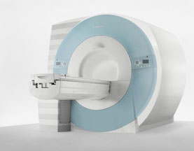
From Siemens Medical Systems;
Received FDA clearance in 2007.
The MAGNETOM Verio provides up to 102 integrated matrix coil elements and up to 32 independent radiofrequency channels that allow flexible coil combinations to make patient and coil repositioning virtually unnecessary. The Tim (total imaging matrix) technology also increases patient throughput due to a shorter scan time.
The open bore design offers great comfort for patients of all shapes and sizes.
Device Information and Specification
CLINICAL APPLICATION
Whole Body
CONFIGURATION
Ultra-short open bore
Head, spine, torso/ body coil, neurovascular, cardiac, neck and multi-purpose flex coils. Peripheral vascular, breast, shoulder, knee, wrist, foot//ankle, TMJ optional.
CHANNELS (min. / max. configuration)
8, 18, 32
Chemical shift imaging, single voxel spectroscopy
MAGNET WEIGHT (gantry included)
8200 kg
DIMENSION H*W*D (gantry included)
173 x 230 x 222 cm
Passive, active; first order,
second order standard
POWER REQUIREMENTS
380 / 400 / 420 / 440 / 460 / 480 V, 3-phase + ground; 110 kVA
| | | | | |
| | | | | |
| |
|
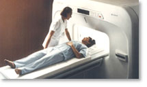
The definition of a scan is to form an image or an electronic representation. The MRI scan uses magnetic resonance principles to produce extremely detailed pictures of the body tissue without the need for X-ray exposure or other damaging forms of radiation.
MRI scans show structures of the different tissues in the body. The tissue that has the least hydrogen atoms (e.g., bones) appears dark, while the tissue with many hydrogen atoms (e.g., fat) looks bright. The MRI pictures of the brain show details and abnormal structures ( brain MRI), for example, tumors, multiple sclerosis lesions, bleedings, or brain tissue that has suffered lack of oxygen after a stroke.
A cardiac MRI scan demonstrates the heart as well as blood vessels ( cardiovascular imaging) and is used to detect heart defects with e.g., changes in the thickness and infarctions of the muscles around the heart. With MRI scans, nearly all kind of body parts can be tested, for example the joints like knee and shoulder, lumbar, thoracic and cervical spine, the pelvis including fetal MRI, and the soft parts of the body such as the liver, kidneys, and spleen.
The MRI procedure includes three to nine imaging sequences and may take up to one hour. See also Lumbar Spine MRI, MRI Safety and Open MRI. | | | | | | | | | | |
• View the DATABASE results for 'MRI Scan' (31).
| | |
• View the NEWS results for 'MRI Scan' (95).
| | | | |  Further Reading: Further Reading: | | Basics:
|
|
News & More:
|  |
A Knee MRI in Half the Time? It's Possible
Thursday, 8 April 2021 by www.diagnosticimaging.com | | |
Michigan radiologist warns about 'incidental findings' in full body MRI scans
Wednesday, 4 October 2023 by www.wilx.com | | |
ACCELERATING MRI SCANS WITH ARTIFICIAL INTELLIGENCE
Friday, 28 August 2020 by www.analyticsinsight.net | | |
Radiographer's Lego Open MRI Product Idea Reaches New Milestone
Monday, 11 November 2019 by www.itnonline.com | | |
Why we need erasable MRI scans
Wednesday, 25 April 2018 by phys.org | | |
MRI as accurate as CT for Crohn's disease detection, management
Tuesday, 6 June 2017 by www.healthimaging.com | | |
MRI scans predict patients' ability to fight the spread of cancer
Tuesday, 12 December 2017 by eurekalert.org | | |
Audio/Video System helps patients relax during MRI scans
Monday, 8 December 2014 by news.thomasnet.com | | |
MRI scans could be a 'game-changer' in prostate cancer testing
Tuesday, 5 August 2014 by www.abc.net.au | | |
7-Tesla MRI scanner allows even more accurate diagnosis of breast cancer
Thursday, 6 March 2014 by www.healthcanal.com |
|
| |
| | | Searchterm 'Knee' was also found in the following services: | | | | |
| | |
| |
|
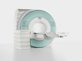
From Hitachi Medical Systems America, Inc.; because of its dependability, the MRP-7000™ remains popular more than a decade after the first U.S. system was shipped. This system maintains a high resale value, what has made it one of the most sought-after scanners on the used MRI equipment market.
Device Information and Specification CLINICAL APPLICATION Whole body DualQuad T/R Body Coil, MA Head, MA C-Spine, MA Shoulder, MA Wrist, MA CTL Spine, MA Knee, MA TMJ, MA Flex Body (3 sizes), Neck, small and large Extremity, PVA (WIP), Breast (WIP), Neurovascular (WIP), Cardiac (WIP) and MA Foot//Ankle (WIP) SE, GE, GR, IR, FIR, STIR, ss-FSE, FSE, DE-FSE/FIR, FLAIR, ss/ms-EPI, ss/ms EPI- DWI, SSP, MTC, SE/GE-EPI, MRCP, SARGE, RSSG, TRSG, BASG, Angiography: CE, PC, 2D/3D TOFIMAGING MODES Single, multislice, volume study horizontal 2.5 m x 2.1 m vertical | | | |
• View the DATABASE results for 'MRP-7000™' (2).
| | | | |
| | | | | |
| |
|


O-scan is manufactured and distributed by Esaote SpA
O-scan is a compact, dedicated extremity MRI system designed for easy installation and high throughput. The complete system fits in a 9' x 10' room, doesn't need for RF or magnetic shielding and it plugs in the wall. The 0.31T permanent magnet along with dual phased array RF coils, and advanced imaging protocols provide outstanding image quality and fast 25 minute complete examinations.
Esaote North America is the exclusive distributor of the O-scan system in the USA.
Device Information and Specification CLINICAL APPLICATION Dedicated Extremity
PULSE SEQUENCES
SE, HSE, HFE, GE, 2dGE, ME, IR, STIR, Stir T2, GESTIR, TSE, TME, FSE STIR, FSE ( T1, T2), X-Bone, Turbo 3DT1, 3D SHARC, 3D SST1, 3D SST2 2D: 2mm - 10 mm, 3D: 0.6 - 10 mm POWER REQUIREMENTS 100/110/200/220/230/240 | | | | | |
| | | | |
| | |
| | | |
|
| |
 | Look
Ups |
| |