Magnetic Resonance - Technology Information Portal Welcome to MRI Technology
Info
  Sheets

Out-
      side


 'Display' 
SEARCH FOR    
 
  2 3 5 A B C D E F G H I J K L M N O P Q R S T U V W X Y Z
Result : Searchterm 'Display' found in 1 term [] and 79 definitions []
previous     36 - 40 (of 80)     next
Result Pages : [1]  [2 3 4 5 6 7 8 9 10 11 12 13 14 15 16]
Searchterm 'Display' was also found in the following services: 
spacer
News  (18)  Resources  (2)  Forum  (11)  
 
FORTE 3.0T™InfoSheet: - Devices -
Intro, 
Types of Magnets, 
Overview, 
etc.MRI Resource Directory:
 - Devices -
 
www.isoltech.co.kr/english/product/30t.htm From ISOL Technology
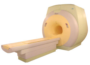
'Ultra high field MR system, it's right close to you. FORTE 3.0T is the new standard for the future ultra high field MR system. If you are pushing the limits of your existing clinical MR scanner, the FORTE will surely take you to the next level of diagnostic imaging. FORTE is the core leader of the medical technology in the 21st century. Proving effects of fMRI that cannot be measured with MRI less than 2.0T.'
Device Information and Specification
CLINICAL APPLICATION
Whole body
CONFIGURATION
Short bore compact
SYNCHRONIZATION
ECG/peripheral: Optional/yes, external trigger, respiratory gating
PULSE SEQUENCES
Spin echo, Gradient echo, Fast spin echo, Inversion recovery, 2D/3D Fast gradient echo sequences FLAIR/STIR, 2D/3D TOF
IMAGING MODES
2D/3D, T1, T2 and Diffusion//Perfusion imaging, MR Angiography package, Advanced EPI package, Multi-nuclei MR Spectroscopy package
FOV
40 cm
128 x 128, 256 x 256, 512 x 512, 1024 x 1024
BORE DIAMETER
or W x H
61 cm without body coil
MAGNET WEIGHT
12000 kg
H*W*D
260 x 220 x 235 cm
COOLING SYSTEM TYPE
Water-cooled coil and air-cooled amplifier
CRYOGEN USE
0.15 L/hr helium
STRENGTH
38 mT/m
5-GAUSS FRINGE FIELD
3.3 m / 5.2 m
Passive and active
spacer
 
• Related Searches:
    • High Field MRI
    • Device
    • Magnet
    • MRI
    • MRI Equipment
MRI Resources 
Brain MRI - Process Analysis - Safety Products - Journals - Spectroscopy pool - Universities
 
Foldover SuppressionInfoSheet: - Sequences - 
Intro, 
Overview, 
Types of, 
etc.
 
A problem occurs in the phase encoding direction, where the phases of signal-bearing tissues outside of the FOV in the y-direction are a replication of the phases that are encoded within the FOV. This signal will be mapped (wrapped, backfolded) back into the image at incorrect locations.
Foldover suppression (phase oversampling, no phase wrap) is a user-selectable parameter that maps this signal to its correct location outside the FOV, then discards any signal from outside the FOV before displaying the image. In order to be able to choose this parameter, in most cases more than an average is necessary.

See also Phase Wrapping Artifact and Oversampling.
spacer

• View the DATABASE results for 'Foldover Suppression' (4).Open this link in a new window

MRI Resources 
Jobs pool - Pregnancy - Distributors - Spectroscopy pool - Cochlear Implant - Nerve Stimulator
 
Hardware
 
MRI hardware includes the electrical and mechanical components of a scanning device.
The main hardware components for the MRI machine are:
The magnet establishing the B0 field to align the spins.
Within the magnet are the gradient coils for producing variations in B0 in the X, Y, and Z directions to make a localization of the received data possible.
Within the gradient coil or directly on the object being imaged is the radio frequency (RF) coil. This RF coil is used to establish the B1 magnetic field necessary to excite the spinning nuclei. The RF coil also detects the signal emitted from the spins within the object being imaged.
The RF amplifier increases the power of the pulses.
The analog to digital converter converts the received analog raw data into digital values.
Depending on the design of the device and the body part being imaged the patient is positioned inside the magnet (e.g. on a movable table or standing upright).
The MRI scan room is surrounded by a RF shield (Faraday cage).
In addition, a computer console, a display, and a film printer belong to the MRI equipment.

See also the related poll result: 'Most outages of your scanning system are caused by failure of'
Radiology-tip.comradCT Scanner,  Gamma Camera
spacer
Medical-Ultrasound-Imaging.comUltrasound Machine
spacer

• View the DATABASE results for 'Hardware' (19).Open this link in a new window


• View the NEWS results for 'Hardware' (1).Open this link in a new window.
 
Further Reading:
  Basics:
Imaging Hardware
   by www.fmrib.ox.ac.uk    
  News & More:
Why non-magnetic capacitors matter in medical imaging
Wednesday, 19 February 2020   by www.medicaldesignandoutsourcing.com    
A transportable MRI machine to speed up the diagnosis and treatment of stroke patients
Wednesday, 22 April 2015   by medicalxpress.com    
Magnetic resonance angiography: current status and future directions
Wednesday, 9 March 2011   by www.jcmr-online.com    
Searchterm 'Display' was also found in the following services: 
spacer
News  (18)  Resources  (2)  Forum  (11)  
 
Infinion 1.5TPanorama 0.2InfoSheet: - Devices -
Intro, 
Types of Magnets, 
Overview, 
etc.MRI Resource Directory:
 - Devices -
 
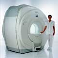
From Philips Medical Systems; www.medical.philips.com/main/products/mri/products/infinion1.5t/features/ Philips Infinion 1.5 T is designed to maximize the efficiency and quality of patient care. Developed with the patient in mind, the Infinion is the shortest and most open 1.5T scanner available. The unique 'ultra short' 1.4 m magnet assures patient comfort and acceptance without compromising image quality and clinical performance.
Device Information and Specification
CLINICAL APPLICATION
Whole body
CONFIGURATION
Ultra short bore
Head, head / neck, integrated C-spine, L/T spine array, small large GP coils, body flex array, torso pelvis array, breast array, endocavitary, shoulder array, lower extremity, hand / wrist, cardiac, PV array
SYNCHRONIZATION
ECG/peripheral, respiratory gating
PULSE SEQUENCES
SE, TSE, SS TSE, EPI, IR, STIR, FLAIR, FFE, TFE, T1 TFE, T2 TFE, Presat, Fatsat, MTC, Diff-opt., Angiography: PCA, MCA, TOF
IMAGING MODES
Single slice, single volume, multi slice, multi volume
TR
3.1 msec minimum
TE
0.9 msec minimum
SINGLE/MULTI SLICE
80 images/sec std.; up to320 opt.@256
FOV
0.4 - 56 cm
2D: 0.3 mm; 3D: 0.2 mm
1024 x 1024
MEASURING MATRIX
8 x 8 to 1024 x 1024
PIXEL INTENSITY
256 gray scale
BORE DIAMETER
or W x H
60 cm diameter (patient)
MAGNET WEIGHT
4100 kg w/cryogen's
H*W*D
233 (lead fitted) x 198 x 140 cm
POWER REQUIREMENTS
400/480 V
COOLING SYSTEM TYPE
Closed loop, chilled water
CRYOGEN USE
0.06 L/hr helium
STRENGTH
30 mT/m
5-GAUSS FRINGE FIELD
3.0 m / 5.0 m
Passive/active
spacer

• View the DATABASE results for 'Infinion 1.5T™' (2).Open this link in a new window

MRI Resources 
Musculoskeletal and Joint MRI - Image Quality - Bioinformatics - Shoulder MRI - Devices - Abdominal Imaging
 
Intera 0.5TPanorama 0.2InfoSheet: - Devices -
Intro, 
Types of Magnets, 
Overview, 
etc.MRI Resource Directory:
 - Devices -
 
www.medical.philips.com/de/products/mri/products/ From Philips Medical Systems;
this low field MRI system of the Intera-family is onto diagnostic safety and flexibility aligned - cost effective and with high patient acceptance.
Device Information and Specification
CLINICAL APPLICATION
Whole body
CONFIGURATION
Short bore compact
Standard: head, body, C1, C3; Optional: Small joint, flex-E, flex-R, endocavitary (L and S), dual TMJ, knee, neck, T/L spine, breast; Optional phased array: Spine, pediatric, 3rd party connector, Flex-S-M-L, flex body, flex cardiac, neuro-vascular, head
SYNCHRONIZATION
ECG/peripheral: Optional/yes, respiratory gating
PULSE SEQUENCES
SE, Modified-SE (TSE), DAVE, STIR, FLAIR, SPIR, MTC, Dynamic, Keyhole, CLEAR, Q Flow, Balanced FFE, Multi Chunk 3D, Multi Stack 3D, FFE-EPI, SE-EPI, IR-EPI, GRASE, Diffusion Imaging, Perfusion Imaging;; Angiography: Inflow MRA, TONE, PCA, CE MRA
IMAGING MODES
Single Slice 2D , Multi Single Slice 2D, Multi Slice 2D, 3D, Multi Chunk 3D, Multi Stack 3D
TR
Min. 2.9 (Omni) msec
TE
Min. 1.0 (Omni) msec
SINGLE/MULTI SLICE
RapidView Recon. greater than 500 @ 256 Matrix
FOV
Max. 53 cm
0.1 mm (Omni)
128 x 128, 256 x 256,512 x 512,1024 x 1024
MEASURING MATRIX
Variable in 1% increments
PIXEL INTENSITY
Lum.: 120 cd/m2; contrast: 150:1
Variable (op. param. depend.)
60 x 60 cm
MAGNET WEIGHT
2500 kg
H*W*D
240 x 188 x 157 cm
POWER REQUIREMENTS
380/400 V
CRYOGEN USE
0.03 L/hr helium
STRENGTH
23 mT/m (Omni)
5-GAUSS FRINGE FIELD
2.1 m / 2.8 m
Passive and dynamic
spacer

• View the DATABASE results for 'Intera 0.5T™' (2).Open this link in a new window

MRI Resources 
MRI Technician and Technologist Schools - Sequences - Open Directory Project - MRI Technician and Technologist Career - Anatomy - MRI Centers
 
previous      36 - 40 (of 80)     next
Result Pages : [1]  [2 3 4 5 6 7 8 9 10 11 12 13 14 15 16]
 Random Page
 
Share This Page
FacebookTwitterLinkedIn

MR-TIP    
Community   
User
Pass
Forgot your UserID/Password ?    


New acceleration techniques will :
reduce scan times 
cause artifacts 
increase expenses 
be useful if you have a lot of experience 
doesn't do much 
never heard of 

Look
      Ups


MR-TIP.com uses cookies! By browsing MR-TIP.com, you agree to our use of cookies.

Magnetic Resonance - Technology Information Portal
Member of SoftWays' Medical Imaging Group - MR-TIP • Radiology-TIP • Medical-Ultrasound-Imaging • 
Copyright © 2003 - 2024 SoftWays. All rights reserved. [ 5 May 2024]
Terms of Use | Privacy Policy | Advertising
 [last update: 2024-02-26 03:41:00]