Magnetic Resonance - Technology Information Portal Welcome to MRI Technology
Info
  Sheets

Out-
      side


 'Reconstruction' 
SEARCH FOR    
 
  2 3 5 A B C D E F G H I J K L M N O P Q R S T U V W X Y Z
Result : Searchterm 'Reconstruction' found in 7 terms [] and 47 definitions []
previous     16 - 20 (of 54)     next
Result Pages : [1 2]  [3 4 5 6 7 8 9 10 11]
Searchterm 'Reconstruction' was also found in the following services: 
spacer
News  (1)  Resources  (3)  Forum  (8)  
 
Asymmetric Sampling
 
The collection of more data points on one side of the k-space origin than on the other. With fewer k-space data points prior to the center (echo) a shorter echo time can be attained. Asymmetric acquisition in any phase encoding direction followed by partial-Fourier reconstruction leads to a reduction in imaging time.
spacer
Searchterm 'Reconstruction' was also found in the following services: 
spacer
Radiology  (22) Open this link in a new windowUltrasound  (6) Open this link in a new window
Aurora® 1.5T Dedicated Breast MRI SystemInfoSheet: - Devices -
Intro, 
Types of Magnets, 
Overview, 
etc.
 
www.auroramri.com/mri/product/ From Aurora Imaging Technology, Inc.;
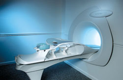
The Aurora® 1.5T Dedicated Breast MRI System with Bilateral SpiralRODEO™ is the first and only FDA approved MRI device designed specifically for breast imaging. The Aurora System, which is already in clinical use at a growing number of leading breast care centers in the US, Europe, got in December 2006 also the approval from the State Food and Drug Administration of the People's Republic of China (SFDA).
'Some of the proprietary and distinguishing features of the Aurora System include: 1) an ellipsoid magnetic shim that provides coverage of both breasts, the chest wall and bilateral axillary lymph nodes; 2) a precision gradient coil with the high linearity required for high resolution spiral reconstruction;; 3) a patient-handling table that provides patient comfort and procedural utility; 4) a fully integrated Interventional System for MRI guided biopsy and localization; and 5) the user-friendly AuroraCAD™ computer-aided image display system designed to improve the accuracy and efficiency of diagnostic interpretations.'
Device Information and Specification
CLINICAL APPLICATION
CONFIGURATION
Short bore compact
IMAGING MODES
Bi-lateral RODEO fat suppression, high resolution Rotating Delivery of Excitation Offresonance Spiral, integrated targeting SW compatible with major MR guided intervention equipment
TR
10 ms for gradient echo and less than 2,500 ms for T2 weighted spin echo
TE
From 5 ms for RODEO Plus to over 80 ms, 120 ms for T2 sequences
SINGLE/MULTI SLICE
Around 0.02 sec for a 256x256 image, 12.4 sec for a 512 x 512 x 32 multislice set
FOV
20 - 36 cm, max. elliptical 36 x 44 cm
MEASURING MATRIX
512 x 512
BORE DIAMETER
or W x H
64 cm diameter (gantry)
MAGNET WEIGHT
8,500 lbs
H*W*D
240 x 188 x 163 cm
POWER REQUIREMENTS
150A/120V-208Y/3 Phase//60 Hz/5 Wire
COOLING SYSTEM
Helium for magnet, distilled/de-ionized water for coil;
spacer

• View the DATABASE results for 'Aurora® 1.5T Dedicated Breast MRI System' (2).Open this link in a new window


• View the NEWS results for 'Aurora® 1.5T Dedicated Breast MRI System' (3).Open this link in a new window.
 
Further Reading:
  News & More:
Aurora Imaging Technology Announces Approval in China
   by salesandmarketingnetwork.com    
Aurora Imaging Technology, Inc. Release: Results of a Multi-Center Trial Demonstrates Superior Diagnostic Performance of the Aurora® 1.5T Dedicated Breast MRI System Over Whole-Body Breast MRI
Monday, 1 October 2012   by www.biospace.com    
MRI Resources 
Veterinary MRI - Hospitals - Case Studies - Chemistry - Spine MRI - Equipment
 
CHORUS 1.5T™InfoSheet: - Devices -
Intro, 
Types of Magnets, 
Overview, 
etc.MRI Resource Directory:
 - Devices -
 
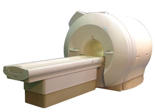
www.isoltech.co.kr/english/product/default.htm 'Next generation MRI system 1.5T CHORUS developed by ISOL Technology is optimized for both clinical diagnostic imaging and for research development.
CHORUS offers the complete range of feature oriented advanced imaging techniques- for both clinical routine and research. The compact short bore magnet, the patient friendly design and the gradient technology make the innovation to new degree of perfection in magnetic resonance.'
Device Information and Specification
CLINICAL APPLICATION
Whole body
CONFIGURATION
Short bore
Head, C-spine, L-spine, TMJ, Knee, Shoulder, General purpose, Phased Array System: 4 digital receiver channels (Up to 12 channels)
Optional
SYNCHRONIZATION
ECG/peripheral: Optional/yes, respiratory gating
PULSE SEQUENCES
Spin Echo, Gradient Echo, Fast Spin Echo, Inversion Recovery (STIR, Fluid Attenuated Inversion Recovery), FLASH, FISP, PSIF, Turbo Flash ( MPRAGE ),TOF MR Angiography, Standard echo planar imaging package (SE-EPI, GE-EPI), Optional: Advanced P.A. Imaging Package (up to 4 ch.), Advanced echo planar imaging package, Single Shot and Diffusion Weighted EPI, IR/FLAIR EPI
IMAGING MODES
2D/3D, Travelling Sat, Multi-Slab 3D, MTC and TONE Pulse Sequence, Fat/Water Suppression, Presaturation (up to 6 bands), Flow Compensation using GMR pulse, Multi-Slice, Multi-Group Imaging
SINGLE/MULTI SLICE
Image reconstruction time (2562 ) : 0.02 s
FOV
40 cm
BORE DIAMETER
or W x H
58 cm diameter (patient)
MAGNET WEIGHT
4050 kg
H*W*D
233 x 206 x 160 cm
COOLING SYSTEM TYPE
Water-cooled coil and air-cooled amplifier
CRYOGEN USE
0.07 L/hr helium
STRENGTH
20 mT/m (Upto 27 mT/m)
5-GAUSS FRINGE FIELD
2.5 m / 3.8 m
Passive and active
spacer

• View the DATABASE results for 'CHORUS 1.5T™' (2).Open this link in a new window

Searchterm 'Reconstruction' was also found in the following services: 
spacer
News  (1)  Resources  (3)  Forum  (8)  
 
Constructive Interference Steady StateInfoSheet: - Sequences - 
Intro, 
Overview, 
Types of, 
etc.
 
(CISS) This gradient echo sequence is a stimulated T2 echo. Two TrueFISP sequences are acquired with differing RF pulses and than combined for strong T2 Weighted high resolution 3D images.
These TrueFISP sequences are normally affected by dark phase dispersion bands, which are caused by patient induced local field inhomogeneities and made prominent by the relatively long TR used. The different excitation pulse regimes offset these bands in the 2 sequences. Combining the images results in a picture free of banding. The image combination is performed automatically after data collection, adding some time to the reconstruction process. The advantage of the 3D CISS sequence is its combination of high signal levels and extremely high spatial resolution.
Used for, e.g. inner ear, cranial nerves and cerebellum.

See also Steady State Free Precession.
spacer
 
Further Reading:
  News & More:
Pediatric and Adult Cochlear Implantation1
2003   by radiographics.rsnajnls.org    
Searchterm 'Reconstruction' was also found in the following services: 
spacer
Radiology  (22) Open this link in a new windowUltrasound  (6) Open this link in a new window
ENCORE 0.5T™InfoSheet: - Devices -
Intro, 
Types of Magnets, 
Overview, 
etc.MRI Resource Directory:
 - Devices -
 
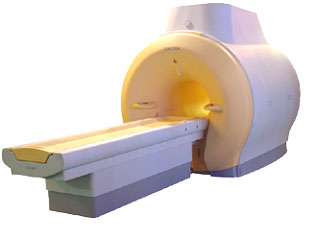
www.isoltech.co.kr/english/product/05t.htm 'MRI system is not an expensive equipment anymore. ENCORE developed by ISOL Technology is a low cost MRI system with the advantages like of the 1.0T MRI scanner. Developed specially for the overseas market, the ENCORE is gaining popularity in the domestic market by medium sized hospitals.
Due to the optimum RF and Gradient application technology. ENCORE enables to obtain high resolution imaging and 2D/3D Angio images which was only possible in high field MR systems.'
- Less consumption of the helium gas due to the ultra-lightweight magnet specially designed and manufactured for ISOL. - Cost efficiency MR system due to air cooling type (equivalent to permanent magnetic). - Patient processing speed of less than 20 minutes.'
Device Information and Specification
CLINICAL APPLICATION
Whole body
CONFIGURATION
Short bore compact
Head, C-spine, L-spine, TMJ, Knee, Shoulder, General purpose, Phased Array System: 4 digital receiver channels (Up to 12 channels)
SYNCHRONIZATION
ECG/peripheral: Optional/yes, respiratory gating
SINGLE/MULTI SLICE
Image reconstruction time (2562 ) : 0.02 s
FOV
40 cm
BORE DIAMETER
or W x H
58 cm diameter
MAGNET WEIGHT
3200 kg
H*W*D
200 x 168 x 187 cm
COOLING SYSTEM TYPE
Air-cooled coil and amplifier
CRYOGEN USE
0.05 L/hr helium
STRENGTH
15 mT/m
5-GAUSS FRINGE FIELD
2.3 m / 3.1 m
Passive and active
spacer

• View the DATABASE results for 'ENCORE 0.5T™' (2).Open this link in a new window

MRI Resources 
Movies - Lung Imaging - Safety pool - Hospitals - Health - Open Directory Project
 
previous      16 - 20 (of 54)     next
Result Pages : [1 2]  [3 4 5 6 7 8 9 10 11]
 Random Page
 
Share This Page
FacebookTwitterLinkedIn

MR-TIP    
Community   
User
Pass
Forgot your UserID/Password ?    


Next big thing in MRI will be :
AI 
remote operator 
personalized protocols 
helium-free 
molecular MRI 
portable MRI 

Look
      Ups


MR-TIP.com uses cookies! By browsing MR-TIP.com, you agree to our use of cookies.

Magnetic Resonance - Technology Information Portal
Member of SoftWays' Medical Imaging Group - MR-TIP • Radiology-TIP • Medical-Ultrasound-Imaging • 
Copyright © 2003 - 2024 SoftWays. All rights reserved. [ 25 April 2024]
Terms of Use | Privacy Policy | Advertising
 [last update: 2024-02-26 03:41:00]