Magnetic Resonance - Technology Information Portal Welcome to MRI Technology
Info
  Sheets

Out-
      side


 'pulse sequences' 
SEARCH FOR    
 
  2 3 5 A B C D E F G H I J K L M N O P Q R S T U V W X Y Z
Result : Searchterm 'pulse sequences' found in 1 term [] and 97 definitions []
previous     36 - 40 (of 98)     next
Result Pages : [1]  [2 3 4 5 6 7 8 9 10 11 12 13 14 15 16 17 18 19 20]
Searchterm 'pulse sequences' was also found in the following services: 
spacer
News  (1)  Resources  (5)  Forum  (8)  
 
G-SCANInfoSheet: - Devices -
Intro, 
Types of Magnets, 
Overview, 
etc.MRI Resource Directory:
 - Devices -
 
www.esaote.de/04_kernspin/gscan/gscan.htm From Esaote S.p.A.; Esaote introduced the new G-SCAN at the RSNA in Dec. 2004. The G-SCAN covers almost all musculoskeletal applications including the spine. The tilting gantry is designed for scanning in weight-bearing positions. This unique MRI scanner is developed in line with the Esaote philosophy of creating high quality MRI systems that are easy to install and that have a low breakeven point.
Device Information and Specification
CLINICAL APPLICATION
CONFIGURATION
Spine, extremity, shoulder, flex coil, knee dual phased array, ankle//foot dual phased array, hand//wrist dual phased array
PULSE SEQUENCES
SE, GE, IR, STIR, TSE, 3D CE, GE-STIR, 3D GE, ME, TME, HSE
IMAGING MODES
Single, multislice, volume study, fast scan, multi slab, cine
FOV
100 up to 350 mm, 25 mm displayed
512 x 512
MEASURING MATRIX
256 x 256 maximum
MAGNET TYPE
Permanent
BORE DIAMETER
or W x H
33 cm H, open
POWER REQUIREMENTS
100/110/200/220/230/240 V
STRENGTH
25 mT/m
5-GAUSS FRINGE FIELD
180 cm
Passive
spacer
MRI Resources 
Examinations - Spine MRI - Online Books - Bioinformatics - Health - Breast Implant
 
Ghosting ArtifactInfoSheet: - Artifacts - 
Case Studies, 
Reduction Index, 
etc.MRI Resource Directory:
 - Artifacts -
 
Quick Overview
Artifact Information
NAME
Ghosting, ghost
DESCRIPTION
Displaced reduplications of image in phase encoding direction
REASON
Motion, heartbeat, respiration
HELP
Triggering, breath hold, pharmaceuticals to reduce bowel motion
Ghosting artifacts are in the most cases caused by movements (e.g., respiratory motion, bowel motion, arterial pulsations, swallowing, and heartbeat) and appear in the phase encoding direction.
mri safety guidance
Image Guidance
Ghosting artifacts can be reduced by respiratory and cardiac triggering, the use of breath holding pulse sequences, flow compensation or presaturation pulses, depending on their origin. To reduce bowel motion also pharmaceuticals, such as glucagon or scopolamine are useful. This will decrease artifacts from both peristalsis and breathing.

See also Motion Artifact, Phase Encoded Motion Artifact, Cardiac Motion Artifact, and Artifact Reduction - Motion.
spacer

• View the DATABASE results for 'Ghosting Artifact' (5).Open this link in a new window

 
Further Reading:
  Basics:
MRI Artifact Gallery
   by chickscope.beckman.uiuc.edu    
MRI Resources 
Developers - Implant and Prosthesis - Spine MRI - Jobs - MR Guided Interventions - Pregnancy
 
Gradient Moment Nulling
 
Gradient moment nulling used as motion artifact suppression technique (MAST) reduces constant velocity motion distortion in standard spin echo or gradient echo pulse sequences. It is an adjustment to zero at the echo time (TE) of the net moments of the amplitude of the waveform of the magnetic field gradients with time. The zeroth moment is the area under the curve. The first moment is the 'center of gravity' etc.
The aim is to minimize the phase shifts acquired by the transverse magnetization of excited nuclei moving along the gradients (including the effect of refocusing radio frequency pulses), particularly for the reduction of image artifacts due to motion.
spacer

• View the DATABASE results for 'Gradient Moment Nulling' (7).Open this link in a new window

 
Further Reading:
  Basics:
Motion Compensation in MR Imaging
   by ccn.ucla.edu    
Searchterm 'pulse sequences' was also found in the following services: 
spacer
News  (1)  Resources  (5)  Forum  (8)  
 
Imagent GIInfoSheet: - Contrast Agents - 
Intro, Overview, 
Characteristics, 
Types of, 
etc.MRI Resource Directory:
 - Contrast Agents -
 
Perflubron® is a perfluorochemical for use as an oral contrast agent. Due to its insolubility in water it does not mix with intestinal secretions; thus bowel lumina appear homogeneously dark on MR images when Perflubron® replaces bowel contents. Filled bowel loops appear black with all pulse sequences because the contrast agent lacks mobile protons.
It is commercially available as Imagent GI. Because rapid transit through the gastrointestinal tract it reaches the rectum within 30 to 40 minutes in most patients. MR imaging of the upper abdominal region should begin within 15 minutes and of the pelvic region 15 to 60 minutes after ingestion of perflubron.

See also Classifications, Characteristics, etc.
Drug Information and Specification
NAME OF COMPOUND
Perfluoroctylbromide
DEVELOPER
CONTRAST EFFECT
Negative enhancement
Proton density reduction, signal void
PHARMACOKINETIC
Gastrointestinal
CONCENTRATION
Water immiscible liquid
DOSAGE
9 mL per kg of body weight
PREPARATION
Finished product
INDICATION
Bowel marking
DEVELOPMENT STAGE
For sale
DISTRIBUTOR
See below
PRESENTATION
Bottle of 200cc
DO NOT RELY ON THE INFORMATION PROVIDED HERE, THEY ARE
NOT A SUBSTITUTE FOR THE ACCOMPANYING PACKAGE INSERT!
Distribution Information
TERRITORY
TRADE NAME
DEVELOPMENT
STAGE
DISTRIBUTOR
USA
Imagent GI®
For sale
spacer

• View the DATABASE results for 'Imagent GI' (3).Open this link in a new window

 
Further Reading:
  News & More:
Slumping MRI market prompts Alliance to halt GI contrast agent effort
Wednesday, 28 September 1994   by www.searchmedica.com    
MRI Resources 
Intraoperative MRI - - PACS - Mobile MRI Rental - Diffusion Weighted Imaging - MR Guided Interventions
 
Infinion 1.5TPanorama 0.2InfoSheet: - Devices -
Intro, 
Types of Magnets, 
Overview, 
etc.MRI Resource Directory:
 - Devices -
 
From Philips Medical Systems; www.medical.philips.com/main/products/mri/products/infinion1.5t/features/ 
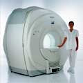
Philips Infinion 1.5 T is designed to maximize the efficiency and quality of patient care. Developed with the patient in mind, the Infinion is the shortest and most open 1.5T scanner available. The unique 'ultra short' 1.4 m magnet assures patient comfort and acceptance without compromising image quality and clinical performance.
Device Information and Specification
CLINICAL APPLICATION
Whole body
CONFIGURATION
Ultra short bore
Head, head / neck, integrated C-spine, L/T spine array, small large GP coils, body flex array, torso pelvis array, breast array, endocavitary, shoulder array, lower extremity, hand / wrist, cardiac, PV array
SYNCHRONIZATION
ECG/peripheral, respiratory gating
PULSE SEQUENCES
SE, TSE, SS TSE, EPI, IR, STIR, FLAIR, FFE, TFE, T1 TFE, T2 TFE, Presat, Fatsat, MTC, Diff-opt., Angiography: PCA, MCA, TOF
IMAGING MODES
Single slice, single volume, multi slice, multi volume
TR
3.1 msec minimum
TE
0.9 msec minimum
SINGLE/MULTI SLICE
80 images/sec std.; up to320 opt.@256
FOV
0.4 - 56 cm
2D: 0.3 mm; 3D: 0.2 mm
1024 x 1024
MEASURING MATRIX
8 x 8 to 1024 x 1024
PIXEL INTENSITY
256 gray scale
BORE DIAMETER
or W x H
60 cm diameter (patient)
MAGNET WEIGHT
4100 kg w/cryogen's
H*W*D
233 (lead fitted) x 198 x 140 cm
POWER REQUIREMENTS
400/480 V
COOLING SYSTEM TYPE
Closed loop, chilled water
CRYOGEN USE
0.06 L/hr helium
STRENGTH
30 mT/m
5-GAUSS FRINGE FIELD
3.0 m / 5.0 m
Passive/active
spacer

• View the DATABASE results for 'Infinion 1.5T™' (2).Open this link in a new window

MRI Resources 
Guidance - Universities - Open Directory Project - Homepages - Databases - Pathology
 
previous      36 - 40 (of 98)     next
Result Pages : [1]  [2 3 4 5 6 7 8 9 10 11 12 13 14 15 16 17 18 19 20]
 Random Page
 
Share This Page
FacebookTwitterLinkedIn

MR-TIP    
Community   
User
Pass
Forgot your UserID/Password ?    


New acceleration techniques will :
reduce scan times 
cause artifacts 
increase expenses 
be useful if you have a lot of experience 
doesn't do much 
never heard of 

Look
      Ups


MR-TIP.com uses cookies! By browsing MR-TIP.com, you agree to our use of cookies.

Magnetic Resonance - Technology Information Portal
Member of SoftWays' Medical Imaging Group - MR-TIP • Radiology-TIP • Medical-Ultrasound-Imaging • 
Copyright © 2003 - 2024 SoftWays. All rights reserved. [ 3 May 2024]
Terms of Use | Privacy Policy | Advertising
 [last update: 2024-02-26 03:41:00]