 | Info
Sheets |
| | | | | | | | | | | | | | | | | | | | | | | | |
 | Out-
side |
| | | | |
|
| | | | | |  | Searchterm 'epi' was also found in the following services: | | | | |
|  |  |
| |
|

(SE) The most common pulse sequence used in MR imaging is based of the detection of a spin or Hahn echo. It uses 90° radio frequency pulses to excite the magnetization and one or more 180° pulses to refocus the spins to generate signal echoes named spin echoes (SE).
In the pulse sequence timing diagram, the simplest form of a spin echo sequence is illustrated.
The 90° excitation pulse rotates the longitudinal magnetization ( Mz) into the xy-plane and the dephasing of the transverse magnetization (Mxy) starts.
The following application of a 180° refocusing pulse (rotates the magnetization in the x-plane) generates signal echoes. The purpose of the 180° pulse is to rephase the spins, causing them to regain coherence and thereby to recover transverse magnetization, producing a spin echo.
The recovery of the z-magnetization occurs with the T1 relaxation time and typically at a much slower rate than the T2-decay, because in general T1 is greater than T2 for living tissues and is in the range of 100-2000 ms.
The SE pulse sequence was devised in the early days of NMR days by Carr and Purcell and exists now in many forms: the multi echo pulse sequence using single or multislice acquisition, the fast spin echo (FSE/TSE) pulse sequence, echo planar imaging ( EPI) pulse sequence and the gradient and spin echo (GRASE) pulse sequence;; all are basically spin echo sequences.
In the simplest form of SE imaging, the pulse sequence has to be repeated as many times as the image has lines. Contrast values:
PD weighted: Short TE (20 ms) and long TR.
T1 weighted: Short TE (10-20 ms) and short TR (300-600 ms)
T2 weighted: Long TE (greater than 60 ms) and long TR (greater than 1600 ms)
With spin echo imaging no T2* occurs, caused by the 180° refocusing pulse. For this reason, spin echo sequences are more robust against e.g., susceptibility artifacts than gradient echo sequences.
See also Pulse Sequence Timing Diagram to find a description of the components.
| | | | | | | | | | | | |  Further Reading: Further Reading: | | Basics:
|
|
News & More:
| |
| |
| | | | | |
| |
|
Argentina-based company, which distributes contrast media like Feridex I.V..
MRI Contrast Agents:
Contact Information MAIL Temis-Lostaló
Zepita 3178
C1285ABF
Ciudad de Buenos Aires
ARGENTINA | | | |
• View the DATABASE results for 'Temis-Lostaló' (3).
| | | | |
| | | | | |
| |
|
In simple ultrafast GRE imaging, TR and TE are so short, that tissues have a poor imaging signal and - more importantly - poor contrast except when contrast media enhanced ( contrast enhanced angiography). Therefore, the magnetization is 'prepared' during the preparation module, most frequently by an initial 180° inversion pulse.
In the pulse sequence timing diagram, the basic ultrafast gradient echo sequence is illustrated. The 180° inversion pulse is executed one time (to the left of the vertical line), the right side represents the data collection period and is often repeated depending on the acquisition parameters.
See also Pulse Sequence Timing Diagram, there you will find a description of the components.
Ultrafast GRE sequences have a short TR,TE, a low flip angle and TR is so short that image acquisition lasts less than 1 second and typically less than 500 ms. Common TR: 3-5 msec, TE: 2 msec, and the flip angle is about 5°.
Such sequences are often labeled with the prefix 'Turbo' like TurboFLASH, TurboFFE and TurboGRASS.
This allows one to center the subsequent ultrafast GRE data acquisition around the inversion time TI, where one of the tissues of interest has very little signal as its z-magnetization is passing through zero.
Unlike a standard inversion recovery (IR) sequence, all lines or a substantial segment of k-space image lines are acquired after a single inversion pulse, which can then together be considered as readout module. The readout module may use a variable flip angle approach, or the data acquisition may be divided into multiple segments (shots). The latter is useful particularly in cardiac imaging where acquiring all lines in a single segment may take too long relative to the cardiac cycle to provide adequate temporal resolution.
If multiple lines are acquired after a single pulse, the pulse sequence is a type of gradient echo echo planar imaging ( EPI) pulse sequence. See also Magnetization Prepared Rapid Gradient Echo ( MPRAGE) and Turbo Field Echo ( TFE). | | | |
• View the DATABASE results for 'Ultrafast Gradient Echo Sequence' (13).
| | | | |
| | | Searchterm 'epi' was also found in the following services: | | | | |
| | |
| |
|
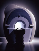
From Toshiba America Medical Systems Inc.;
With its high-field strength, the Vantage™ delivers the clinical capabilities and image quality expected by cardiologists, while simultaneously offering patients a more comfortable and non-invasive option, said Dane Peshe, director, MRI Business Unit, Toshiba America Medical Systems. Vantage™ supports a full complement of cardiovascular imaging studies, ranging from stroke evaluation to peripheral vascular imaging. Additionally, the ultra short bore design offers patients a greater feeling of openness that reduces claustrophobic sensations, while Toshiba's exclusive, patented Pianissimo™ technology reduces scan noise by as much as 90 percent for a more pleasant experience.'
Device Information and Specification CLINICAL APPLICATION Whole body CONFIGURATION Ultra short bore SE, FE, IR, FastSE, FastIR, FastFLAIR, Fast STIR, FastFE, FASE, EPI, SuperFASE; Angiography: 2D(gate/non-gate)/3D TOF, SORS-STC IMAGING MODES Single, multislice, volume study 32-1024, phase;; 64-1024, freq. POWER REQUIREMENTS 380/400/415/440/480 V COOLING SYSTEM TYPE Closed-loop water-cooled Liquid helium: approx. less than 0.05 L/hr Passive, active, auto-active | | | |
• View the DATABASE results for 'Vantage™' (2).
| | | | | | Further Reading: | Basics:
|
|
| |
| | | | | | | | | |
| | |
| | | |
|
| |
 | Look
Ups |
| |


