Magnetic Resonance - Technology Information Portal Welcome to MRI Technology
Info
  Sheets

Out-
      side


 'Ultrafast Spin Echo' 
SEARCH FOR    
 
  2 3 5 A B C D E F G H I J K L M N O P Q R S T U V W X Y Z
Result : Searchterm 'Ultrafast Spin Echo' found in 0 term [] and 0 definition [], (+ 3 Boolean[] results
1 - 3 (of 3)     
Result Pages : [1]
Searchterm 'Ultrafast Spin Echo' was also found in the following service: 
spacer
News  (1)  
 
Gradient Echo SequenceForum -
related threadsInfoSheet: - Sequences - 
Intro, 
Overview, 
Types of, 
etc.
 
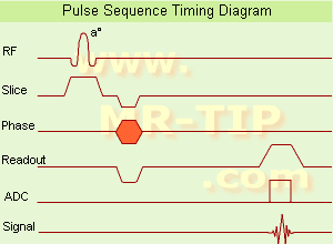
Gradient Echo Sequence Timing Diagram (GRE - sequence) A gradient echo is generated by using a pair of bipolar gradient pulses. In the pulse sequence timing diagram, the basic gradient echo sequence is illustrated. There is no refocusing 180° pulse and the data are sampled during a gradient echo, which is achieved by dephasing the spins with a negatively pulsed gradient before they are rephased by an opposite gradient with opposite polarity to generate the echo.
See also the Pulse Sequence Timing Diagram. There you will find a description of the components.
The excitation pulse is termed the alpha pulse α. It tilts the magnetization by a flip angle α, which is typically between 0° and 90°. With a small flip angle there is a reduction in the value of transverse magnetization that will affect subsequent RF pulses. The flip angle can also be slowly increased during data acquisition (variable flip angle: tilt optimized nonsaturation excitation). The data are not acquired in a steady state, where z-magnetization recovery and destruction by ad-pulses are balanced. However, the z-magnetization is used up by tilting a little more of the remaining z-magnetization into the xy-plane for each acquired imaging line.
Gradient echo imaging is typically accomplished by examining the FID, whereas the read gradient is turned on for localization of the signal in the readout direction. T2* is the characteristic decay time constant associated with the FID. The contrast and signal generated by a gradient echo depend on the size of the longitudinal magnetization and the flip angle. When α = 90° the sequence is identical to the so-called partial saturation or saturation recovery pulse sequence. In standard GRE imaging, this basic pulse sequence is repeated as many times as image lines have to be acquired. Additional gradients or radio frequency pulses are introduced with the aim to spoil to refocus the xy-magnetization at the moment when the spin system is subject to the next α pulse.
As a result of the short repetition time, the z-magnetization cannot fully recover and after a few initial α pulses there is an equilibrium established between z-magnetization recovery and z-magnetization reduction due to the α pulses.
Gradient echoes have a lower SAR, are more sensitive to field inhomogeneities and have a reduced crosstalk, so that a small or no slice gap can be used. In or out of phase imaging depending on the selected TE (and field strength of the magnet) is possible. As the flip angle is decreased, T1 weighting can be maintained by reducing the TR. T2* weighting can be minimized by keeping the TE as short as possible, but pure T2 weighting is not possible. By using a reduced flip angle, some of the magnetization value remains longitudinal (less time needed to achieve full recovery) and for a certain T1 and TR, there exist one flip angle that will give the most signal, known as the "Ernst angle".
Contrast values:
PD weighted: Small flip angle (no T1), long TR (no T1) and short TE (no T2*)
T1 weighted: Large flip angle (70°), short TR (less than 50ms) and short TE
T2* weighted: Small flip angle, some longer TR (100 ms) and long TE (20 ms)

Classification of GRE sequences can be made into four categories:
See also Gradient Recalled Echo Sequence, Spoiled Gradient Echo Sequence, Refocused Gradient Echo Sequence, Ultrafast Gradient Echo Sequence.
 
Images, Movies, Sliders:
 MRI Liver In Phase  Open this link in a new window
    
 MRI Liver Out Of Phase  Open this link in a new window
    
 MVP Parasternal  Open this link in a new window
 Breast MRI Images T1 Pre - Post Contrast  Open this link in a new window
 Circle of Willis, Time of Flight, MIP  Open this link in a new window
    
SlidersSliders Overview

 
spacer
 
• Share the entry 'Gradient Echo Sequence':  Facebook  Twitter  LinkedIn  
 
• Related Searches:
    • Ultrafast Gradient Echo Sequence
    • MRI History
    • Abdominal Imaging
    • Black Boundary Artifact
    • Pulse Sequence Timing Diagram
 
Further Reading:
  Basics:
Enhanced Fast GRadient Echo 3-Dimensional (efgre3D) or THRIVE
   by www.mri.tju.edu    
  News & More:
MRI evaluation of fatty liver in day to day practice: Quantitative and qualitative methods
Wednesday, 3 September 2014   by www.sciencedirect.com    
T1rho-prepared balanced gradient echo for rapid 3D T1rho MRI
Monday, 1 September 2008   by www.ncbi.nlm.nih.gov    
MRI Resources 
Brain MRI - Resources - Functional MRI - Mobile MRI Rental - MRI Reimbursement - Portals
 
Fetal MRI
 
Ultrasound imaging is the primary fetal monitoring modality during pregnancy, nevertheless fetal MRI is increasingly used to image anatomical regions and structures difficult to see with sonography. Given its long record of safety, utility, and cost-effectiveness, ultrasound will remain the modality of first choice in fetal screening. However, MRI is beginning to fill a niche in situations where ultrasound does not provide enough information to diagnose abnormalities before the baby's birth. Magnetic resonance imaging of the fetus provides multiplanar views also in sub-optimal positions, better characterization of anatomic details of e.g. the fetal brain, and information for planning the mode of delivery and airway management at birth.

Indications:
Fetal anomalies
Maternal tumors
Pelvimetry
Examinations of the placenta

Modern fetal MRI requires no sedatives or muscle relaxants to control fetal movement. Ultrafast MRI techniques (e.g., single shot techniques like Half Fourier Acquisition Single shot Turbo spin Echo HASTE) enable images to be acquired in less than one second to eliminate fetal motion. Such technology has led to increased usage of fetal MRI, which can lead to earlier diagnosis of conditions affecting the baby and has proven useful in planning fetal surgery and designing postnatal treatments. As MR technology continues to improve, more advances in the prenatal diagnosis and treatment of fetal abnormalities are to expect. More advances in in-utero interventions are likely as well. Eventually, fetal MRI may replace even some prenatal tests that require invasive procedures such as amniocentesis.

For Ultrasound Imaging (USI) see Fetal Ultrasound at Medical-Ultrasound-Imaging.com.
 
Images, Movies, Sliders:
 Normal Fetus  Open this link in a new window
    

Courtesy of  Robert R. Edelman
 Pregnancy and Small Bowel Obstruction  Open this link in a new window
    

Courtesy of  Robert R. Edelman
 Fetus (Brain) and Dermoid in Mother  Open this link in a new window
      

Courtesy of  Robert R. Edelman

 
spacer

• View the DATABASE results for 'Fetal MRI' (5).Open this link in a new window


• View the NEWS results for 'Fetal MRI' (2).Open this link in a new window.
 
Further Reading:
  Basics:
Fetal MRI is a Valuable Adjunct to Ultrasound in Detecting Abnormal Extracardiac Development in Fetuses with Congenital Heart Defects
Friday, 24 December 2021   by www.itnonline.com    
Specific Absorption Rate and Specific Energy Dose: Comparison of 1.5-T versus 3.0-T Fetal MRI
Tuesday, 7 April 2020   by pubs.rsna.org    
Untangling the Maze, Imaging the Fetus
Tuesday, 30 September 2014   by www.newswise.com    
In fetal MRI, 3T shown to have it all over 1.5T
Tuesday, 12 January 2016   by www.healthimaging.com    
  News & More:
Advances in medical imaging enable visualization of white matter tracts in fetuses
Wednesday, 12 May 2021   by www.eurekalert.or    
Fetal CMR Detects Congenital Heart Defects, Changes Treatment Decisions
Monday, 29 March 2021   by www.diagnosticimaging.com    
MRI scans more precisely define and detect some abnormalities in unborn babies
Friday, 12 March 2021   by www.eurekalert.org    
Ultrasound and Magnetic Resonance Imaging of Agenesis of the Corpus Callosum in Fetuses: Frontal Horns and Cavum Septi Pellucidi Are Clues to Earlier Diagnosis
Monday, 29 June 2020   by pubmed.ncbi.nlm.nih.gov    
MRI helps predict preterm birth
Tuesday, 15 March 2016   by www.eurekalert.org    
3-T MRI advancing on ultrasound for imaging fetal abnormalities
Monday, 20 April 2015   by www.eurekalert.org    
Babies benefit from pioneering 'miniature' MRI scanner in Sheffield
Friday, 24 January 2014   by www.telegraph.co.uk    
Ultrasensitive Detector Pinpoints Big Problem in Tiny Fetal Heart
Tuesday, 6 April 2010   by www.sciencedaily.com    
Real-time MRI helps doctors assess beating heart in fetus
Thursday, 29 September 2005   by www.eurekalert.org    
MRI Resources 
Musculoskeletal and Joint MRI - Veterinary MRI - Movies - Directories - Liver Imaging - RIS
 
Cardiac Gating
 
This method synchronize the heartbeat with the beginning of the TR, whereat the r wave is used as the trigger. Cardiac gating times the acquisition of MR data to physiological motion in order to minimize motion artifacts. ECG gating techniques are useful whenever data acquisition is too slow to occur during a short fraction of the cardiac cycle.
Image blurring due to cardiac-induced motion occurs for imaging times of above approximately 50 ms in systole, while for imaging during diastole the critical time is of the order of 200-300 ms. The acquisition of an entire image in this time is only possible with using ultrafast MR imaging techniques. If a series of images using cardiac gating or real-time echo planar imaging EPI are acquired over the entire cardiac cycle, pixel-wise velocity and vascular flow can be obtained.
In simple cardiac gating, a single image line is acquired in each cardiac cycle. Lines for multiple images can then be acquired successively in consecutive gate intervals. By using the standard multiple slice imaging and a spin echo pulse sequence, a number of slices at different anatomical levels is obtained. The repetition time (TR) during a ECG-gated acquisition equals the RR interval, and the RR interval defines the minimum possible repetition time (TR). If longer TRs are required, multiple integers of the RR interval can be selected. When using a gradient echo pulse sequence, multiple phases of a single anatomical level or multiple slices at different anatomical levels can be acquired over the cardiac cycle.
Also called cardiac triggering.
 
Images, Movies, Sliders:
 Cardiac Infarct Short Axis Cine Overview  Open this link in a new window
    

Courtesy of  Robert R. Edelman
 Infarct 4 Chamber Cine  Open this link in a new window
    

Courtesy of  Robert R. Edelman
 
spacer

• View the DATABASE results for 'Cardiac Gating' (15).Open this link in a new window

 
Further Reading:
  Basics:
Cardiac MRI - Technical Aspects Primer
Wednesday, 7 August 2002
Electrocardiogram in an MRI Environment: Clinical Needs, Practical Considerations, Safety Implications, Technical Solutions and Future Directions
Wednesday, 25 January 2012   by cdn.intechopen.com    
Motion-compensation of Cardiac Perfusion MRI using a Statistical Texture Ensemble(.pdf)
June 2003   by www.imm.dtu.dk    
Searchterm 'Ultrafast Spin Echo' was also found in the following service: 
spacer
News  (1)  
 
     1 - 3 (of 3)     
Result Pages : [1]
 Random Page
 
Share This Page
FacebookTwitterLinkedIn

MR-TIP    
Community   
User
Pass
Forgot your UserID/Password ?    


Personalized protocols (age, gender, body habitus, etc.) lead to :
more automated planning 
improved patient comfort 
better diagnostics 
optimized image quality 
nothing 

Look
      Ups


MR-TIP.com uses cookies! By browsing MR-TIP.com, you agree to our use of cookies.

Magnetic Resonance - Technology Information Portal
Member of SoftWays' Medical Imaging Group - MR-TIP • Radiology-TIP • Medical-Ultrasound-Imaging • 
Copyright © 2003 - 2024 SoftWays. All rights reserved. [ 20 May 2024]
Terms of Use | Privacy Policy | Advertising
 [last update: 2024-02-26 03:41:00]