 | Info
Sheets |
| | | | | | | | | | | | | | | | | | | | | | | | |
 | Out-
side |
| | | | |
|
| | | | |
Result : Searchterm 'R1' found in 1 term [ ] and 39 definitions [ ] and 39 definitions [ ] ]
| | previous 36 - 40 (of 40) Result Pages : [1] [2 3 4 5 6 7 8] |  | |  | Searchterm 'R1' was also found in the following services: | | | | |
| |  |
| |
|
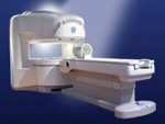
From GE Healthcare;
the Signa Ovation™ is a patient-friendly open MRI scanner designed not only to handle a typical patient mix, but to accommodate larger patients, patients who are claustrophobic, and others who have difficulty tolerating the close quarters of conventional MR machines.
Device Information and Specification CLINICAL APPLICATION Whole body Standard: SE, IR, 2D/3D GRE and SPGR, 2D/3D TOF, 2D/3D FSE, 2D/3D FGRE and FSPGR, SSFP, FLAIR, EPI, optional: 2D/3D Fiesta, true chem sat, fat/water separation, single shot diffusion EPI, line scan diffusionIMAGING MODES Localizer, single slice, multislice, volume, fast, POMP, multi slab, cine, slice and frequency zip, extended dynamic range, tailored RF TR 1.3 to 12000 msec in increments of 1 msec TE 0.4 to 2000 msec in increments of 1 msec 2D: 1.4mm - 20mm 3D: 0.2mm - 20mm 0.08 mm; 0.02 mm optional POWER REQUIREMENTS 200 - 480, 3-phase MAX. GRADIENT AMPLITUDE 19 mT/m | | | | | |
| | | Searchterm 'R1' was also found in the following service: | | | | |
| | |
| |
|

From GE Healthcare;
The Signa SP 0.5T™ is an open MRI magnet that is designed for use in interventional radiology and intra-operative imaging. The vertical gap configuration increases patient positioning options, improves patient observation, and allows continuous access to the patient during imaging.
The magnet enclosure also incorporates an intercom, patient observation video camera, laser patient alignment lights, and task lighting in the imaging volume.
Device Information and Specification CLINICAL APPLICATION Whole body Integrated transmit and receive body coil; optional rotational body coil, head; other coils optional; open architecture makes system compatible with a wide selection of coilsarray Standard: SE, IR, 2D/3D GRE and SPGR, 2D/3D TOF, 2D/3D FSE, 2D/3D FGRE and FSPGR, SSFP, FLAIR, EPI, optional: 2D/3D Fiesta, true chem sat, fat/water separation, single shot diffusion EPI IMAGING MODES Localizer, single slice, multislice, volume, fast, POMP, multi slab, cine, slice and frequency zip, extended dynamic range, tailored RF TR 1.3 to 12000 msec in increments of 1 msec TE 0.4 to 2000 msec in increments of 1 msec 2D: 1.4mm - 20mm 3D: 0.2mm - 20mm POWER REQUIREMENTS 200 - 480, 3-phase | | | |
• View the DATABASE results for 'Signa SP 0.5T™ Open Configuration' (2).
| | | | |  Further Reading: Further Reading: | News & More:
|
|
| |
| | | | | |
| |
|
(Mn-DPDP) This agent, mangafodipir trisodium, is a hepatocyte specific MRI contrast agent. Manganese is very toxic, so it has to be chelated and put in the form of a vitamin B6 analog, which is taken up by normal hepatocytes to some extent.
Teslascan® was developed in the early 1980's, went through clinical trials in the early 1990's, and was approved in 1997. One problem with assessing the efficacy of this agent is the fact that the phase III trials finished in the early 1990's, and the techniques used for MR today are very different from the techniques used almost a decade ago.
This contrast agent shortens the T1 relaxation time. On T1 weighted pictures it makes a normal liver look brighter. Since metastases, for example, do not generally take up this agent, the contrast between the enhancing liver and the non-enhancing lesions will increase on T1 weighted pictures. It does not have much effect on T2 weighted images. Drug Information and Specification T1, Predominantly positive enhancement PHARMACOKINETIC Hepatobiliary, pancreatic, adrenal DOSAGE 5 µmol/kg, 0.5 ml/kg PREPARATION Finished product DEVELOPMENT STAGE Approved PRESENTATION Vials of 100 ml DO NOT RELY ON THE INFORMATION PROVIDED HERE, THEY ARE
NOT A SUBSTITUTE FOR THE ACCOMPANYING PACKAGE INSERT! Distribution Information TERRITORY TRADE NAME DEVELOPMENT
STAGE DISTRIBUTOR | | | |
• View the DATABASE results for 'Teslascan®' (4).
| | | | | | Further Reading: | | Basics:
|
|
News & More:
| |
| |
| | | Searchterm 'R1' was also found in the following services: | | | | |
| | |
| |
|
(VSOP = very small superparamagnetic iron oxide particles) This new class of contrast agents with smaller particle size than SPIO offers advantages for MR angiography. SPIO particles are usually coated with an organic polymer such as dextran, carboxydextran or polyethylene glycol, which limits the minimal overall particle size that can be obtained.
VSOP-C184 consists of an aqueous solution of superparamagnetic iron oxide particles with a citrate coating and the overall particle size of 4-8 nm.
Short name: VSOP-C184, central moiety: Fe, concentration: 29 g Fe/l, relaxivity: R1=20.1, R2=37.1, B0=0.94 T. | | | | | |
| | | Searchterm 'R1' was also found in the following service: | | | | |
| | |
| |
|
Short name: WIN 22181, central moiety: Gd2+, relaxivity: r1 = 9.5
A substance under development (preclin.) as a contrast agent for MR urography. | | | | | |
| | | | |
| | |
| | | |
|
| |
 | Look
Ups |
| |


