Magnetic Resonance - Technology Information Portal Welcome to MRI Technology
Info
  Sheets

Out-
      side


 'Pulse Length' 
SEARCH FOR    
 
  2 3 5 A B C D E F G H I J K L M N O P Q R S T U V W X Y Z
Result : Searchterm 'Pulse Length' found in 1 term [] and 1 definition [], (+ 6 Boolean[] results
1 - 5 (of 8)     next
Result Pages : [1]  [2]
MRI Resources 
Manufacturers - Corporations - Examinations - Raman Spectroscopy - Pathology - Jobs
 
Pulse Length
 
Time duration of a pulse. For an RF pulse near the Larmor frequency, the longer the pulse length, the greater the angle of rotation of the macroscopic magnetization vector will be (greater than 180° can bring it back toward its original orientation). For an RF pulse of a given shape as a function of time, the longer the pulse length, the narrower the equivalent range of frequencies in the pulse will be.
spacer
 
• Share the entry 'Pulse Length':  Facebook  Twitter  LinkedIn  
 
Further Reading:
  News & More:
Factors influencing flip angle mapping in MRI: RF pulse shape, slice-select gradients, off-resonance excitation, and B0 inhomogeneities.
Tuesday, 1 August 2006   by www.ncbi.nlm.nih.gov    
Searchterm 'Pulse Length' was also found in the following service: 
spacer
Ultrasound  (2) Open this link in a new window
Pulse Width
 
spacer
MRI Resources 
MRI Technician and Technologist Career - Pacemaker - Claustrophobia - Patient Information - Societies - Diffusion Weighted Imaging
 
Fast Spin EchoForum -
related threadsInfoSheet: - Sequences - 
Intro, 
Overview, 
Types of, 
etc.
 
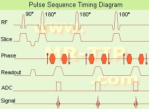
Fast Spin Echo Diagram (FSE) In the pulse sequence timing diagram, a fast spin echo sequence with an echo train length of 3 is illustrated. This sequence is characterized by a series of rapidly applied 180° rephasing pulses and multiple echoes, changing the phase encoding gradient for each echo.
The echo time TE may vary from echo to echo in the echo train. The echoes in the center of the K-space (in the case of linear k-space acquisition) mainly produce the type of image contrast, whereas the periphery of K-space determines the spatial resolution. For example, in the middle of K-space the late echoes of T2 weighted images are encoded. T1 or PD contrast is produced from the early echoes.
The benefit of this technique is that the scan duration with, e.g. a turbo spin echo turbo factor / echo train length of 9, is one ninth of the time. In T1 weighted and proton density weighted sequences, there is a limit to how large the ETL can be (e.g. a usual ETL for T1 weighted images is between 3 and 7). The use of large echo train lengths with short TE results in blurring and loss of contrast. For this reason, T2 weighted imaging profits most from this technique.
In T2 weighted FSE images, both water and fat are hyperintense. This is because the succession of 180° RF pulses reduces the spin spin interactions in fat and increases its T2 decay time. Fast spin echo (FSE) sequences have replaced conventional T2 weighted spin echo sequences for most clinical applications. Fast spin echo allows reduced acquisition times and enables T2 weighted breath hold imaging, e.g. for applications in the upper abdomen.
In case of the acquisition of 2 echoes this type of a sequence is named double fast spin echo / dual echo sequence, the first echo is usually density and the second echo is T2 weighted image. Fast spin echo images are more T2 weighted, which makes it difficult to obtain true proton density weighted images. For dual echo imaging with density weighting, the TR should be kept between 2000 - 2400 msec with a short ETL (e.g., 4).
Other terms for this technique are:
Turbo Spin Echo
Rapid Imaging Spin Echo,
Rapid Spin Echo,
Rapid Acquisition Spin Echo,
Rapid Acquisition with Refocused Echoes
 
Images, Movies, Sliders:
 Lumbar Spine T2 FSE Sagittal  Open this link in a new window
    

Courtesy of  Robert R. Edelman
 MRI - Anatomic Imaging of the Foot  Open this link in a new window
    
SlidersSliders Overview

 Lumbar Spine T2 FSE Axial  Open this link in a new window
    

Courtesy of  Robert R. Edelman
 
spacer

• View the DATABASE results for 'Fast Spin Echo' (31).Open this link in a new window

 
Further Reading:
  Basics:
MYELIN-SELECTIVE MRI: PULSE SEQUENCE DESIGN AND OPTIMIZATION
   by www.imaging.robarts.ca    
Advances in Magnetic Resonance Neuroimaging
Friday, 27 February 2009   by www.ncbi.nlm.nih.gov    
  News & More:
New MR sequence helps radiologists more accurately evaluate abnormalities of the uterus and ovaries
Thursday, 23 April 2009   by www.eurekalert.org    
Spin echoes, CPMG and T2 relaxation - Introductory NMR & MRI from Magritek
2013   by www.azom.com    
MRI Resources 
Nerve Stimulator - Equipment - MRI Training Courses - Manufacturers - Absorption and Emission - Fluorescence
 
Double Inversion Recovery T1 MeasurementInfoSheet: - Sequences - 
Intro, 
Overview, 
Types of, 
etc.MRI Resource Directory:
 - Sequences -
 
(DIR or DIRT1) Double inversion recovery T1 measurement is a T1 weighted black blood MRA sequence in which the signal from blood is suppressed. The inversion time to suppress blood is described as the duration between the initial inversion pulse and time point that the longitudinal magnetization of blood reaches the zero point. The readout starts at the blood suppression inversion time (BSP TI) and blood in the imaging slice gives no signal. This inversion time is around 650 ms with a 60 beat per minute heart rate at 1.5 T.
The TI can be decreased by using a wider receive bandwidth, shorter echo train length and/or narrow trigger window. Wide bandwidth also decreases the blurring caused by long echo trains at the expense of signal to noise ratio. In case of in plane or slow flow the suppression of the signal from blood may be incomplete. With increased TE or change of the image plane the blood suppression can be improved.
Double inversion recovery is a breath hold technique with one image per acquisition used in cardiovascular imaging. The patient is instructed to hold the breath in expiration (if not possible also inspiration can be taken), so that the end diastolic volume in the cardiac chambers would be the same during entire scanning. DIR provides fine details of the boundary between the lumen and the wall of the cardiac chambers and main vascular and heart structures, pericardium, and mediastinal tissues.
 
Images, Movies, Sliders:
 Normal Dual Inversion Fast Spin-echo  Open this link in a new window
      

Courtesy of  Robert R. Edelman

 
spacer

• View the DATABASE results for 'Double Inversion Recovery T1 Measurement' (2).Open this link in a new window

 
Further Reading:
  News & More:
Artificial double inversion recovery images can substitute conventionally acquired images: an MRI-histology study
Wednesday, 16 February 2022   by www.nature.com    
Searchterm 'Pulse Length' was also found in the following service: 
spacer
Ultrasound  (2) Open this link in a new window
Generalized Autocalibrating Partially Parallel AcquisitionInfoSheet: - Sequences - 
Intro, 
Overview, 
Types of, 
etc.
 
(GRAPPA) GRAPPA is a parallel imaging technique to speed up MRI pulse sequences. The Fourier plane of the image is reconstructed from the frequency signals of each coil (reconstruction in the frequency domain).
Parallel imaging techniques like GRAPPA, auto-SMASH and VD-AUTO-SMASH are second and third generation algorithms using k-space undersampling. A model from a part of the center of k-space is acquired, to find the coefficients of the signals from each coil element, and to reconstruct the missing intermediary lines. The acquisition of these additional lines is a form of self-calibration, which lengthens the overall short scan time. The acquisition of these k-space lines provides mapping of the whole field as well as data for the image contrast.
Algorithms of the GRAPPA type work better than the SENSE type in heterogeneous body parts like thoracic or abdominal imaging, or in pulse sequences like echo planar imaging. This is caused by differences between the sensitivity map and the pulse sequence (e.g. artifacts) or an unreliable sensitivity map.
spacer

• View the DATABASE results for 'Generalized Autocalibrating Partially Parallel Acquisition' (2).Open this link in a new window

MRI Resources 
MRI Reimbursement - Homepages - Brain MRI - Corporations - Used and Refurbished MRI Equipment - Safety pool
 
     1 - 5 (of 8)     next
Result Pages : [1]  [2]
 Random Page
 
Share This Page
FacebookTwitterLinkedIn

MR-TIP    
Community   
User
Pass
Forgot your UserID/Password ?    


Next big thing in MRI will be :
AI 
remote operator 
personalized protocols 
helium-free 
molecular MRI 
portable MRI 

Look
      Ups


MR-TIP.com uses cookies! By browsing MR-TIP.com, you agree to our use of cookies.

Magnetic Resonance - Technology Information Portal
Member of SoftWays' Medical Imaging Group - MR-TIP • Radiology-TIP • Medical-Ultrasound-Imaging • 
Copyright © 2003 - 2024 SoftWays. All rights reserved. [ 26 April 2024]
Terms of Use | Privacy Policy | Advertising
 [last update: 2024-02-26 03:41:00]