 | Info
Sheets |
| | | | | | | | | | | | | | | | | | | | | | | | |
 | Out-
side |
| | | | |
|
| | | | | |  | Searchterm 'Mode' was also found in the following services: | | | | |
|  |  |
| |
|

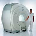
The company is a leading manufacturer and developer of magnetic resonance imaging ( MRI) scanners.
The Patient Friendly MRI Company, formed in 1978, is engaged in the business of inventing, manufacturing, selling and servicing magnetic resonance imaging ( MRI) scanners. FONAR is the oldest MRI company in the world. After receiving hundreds of millions in a windfall from protecting their MRI patents, they made a MRI scanner that no other MRI manufacturer has. One that the patient stands in and they call Indomitable, the Stand-Up MRI. Patients like it because it is the least claustrophobic, most comfortable MRI on the market. Doctors like it because of its superior image quality and for the first time, the patient can be scanned in the weight-bearing position, or the position of pain or symptom. In October of 2004, the company changed the product name of the Stand-Up MRI to the Upright MRI. Fonar introduced the first "open" MRI scanner in 1980 and is the originator of the iron-core nonsuperconductive and permanent magnet technology.
MRI Scanners:
- 0.6T:
•
QUAD™ 12000 - Its 19-inch gap and Whisper Gradients™ make it extraordinarily spacious, quiet and comfortable. With its signal to noise advantage of 0.6 T and its comprehensive array of Organ-Specific™ receiver coils, the QUAD™ 12000 provides high-speed, high resolution and high contrast scanning.
Product Specification
•
OR 360°™ - cleared for marketing by the FDA in March 2000, 360° access to the patient. A dual-purpose scanner, it can be used for conventional diagnostic scanning when not in surgical mode.
Product Specification
Contact Information
MAIL
FONAR Corporation
110 Marcus Drive
Melville, N.Y. 11747
USA
| | | |
• View the NEWS results for 'FONAR Corporation' (87).
| | | | |  Further Reading: Further Reading: | | Basics:
|
|
News & More:
| |
| |
| | | | | |
| |
|

From ISOL Technology
'Ultra high field MR system, it's right close to you.
FORTE 3.0T is the new standard for the future ultra high field MR system.
If you are pushing the limits of your existing clinical MR scanner, the FORTE will surely take you to the next level of diagnostic imaging.
FORTE is the core leader of the medical technology in the 21st century. Proving effects of fMRI that cannot be measured with MRI less than 2.0T.'
Device Information and Specification
CLINICAL APPLICATION
Whole body
CONFIGURATION
Short bore compact
128 x 128, 256 x 256, 512 x 512, 1024 x 1024
| | | |
• View the DATABASE results for 'FORTE 3.0T™' (2).
| | | | |
| | | | | |
| |
|

From Esaote S.p.A.;
Esaote introduced the new G-SCAN at the RSNA in Dec. 2004. The G-SCAN covers almost all musculoskeletal applications including the spine. The tilting gantry is designed for scanning in weight-bearing positions. This unique MRI scanner is developed in line with the Esaote philosophy of creating high quality MRI systems that are easy to install and that have a low breakeven point.
Device Information and Specification
SE, GE, IR, STIR, TSE, 3D CE, GE-STIR, 3D GE, ME, TME, HSE
100 up to 350 mm, 25 mm displayed
POWER REQUIREMENTS
100/110/200/220/230/240 V
| | | |
• View the DATABASE results for 'G-SCAN' (3).
| | | | |
| | | Searchterm 'Mode' was also found in the following services: | | | | |
| | |
| |
|
(GRAPPA) GRAPPA is a parallel imaging technique to speed up MRI pulse sequences. The Fourier plane of the image is reconstructed from the frequency signals of each coil ( reconstruction in the frequency domain).
Parallel imaging techniques like GRAPPA, auto-SMASH and VD-AUTO-SMASH are second and third generation algorithms using k-space undersampling. A model from a part of the center of k-space is acquired, to find the coefficients of the signals from each coil element, and to reconstruct the missing intermediary lines. The acquisition of these additional lines is a form of self-calibration, which lengthens the overall short scan time. The acquisition of these k-space lines provides mapping of the whole field as well as data for the image contrast.
Algorithms of the GRAPPA type work better than the SENSE type in heterogeneous body parts like thoracic or abdominal imaging, or in pulse sequences like echo planar imaging. This is caused by differences between the sensitivity map and the pulse sequence (e.g. artifacts) or an unreliable sensitivity map. | | | |
• View the DATABASE results for 'Generalized Autocalibrating Partially Parallel Acquisition' (2).
| | | | |
| | | | | |
| |
|
From Philips Medical Systems;
Philips Infinion 1.5 T is designed to maximize the efficiency and quality of patient care. Developed with the patient in mind, the Infinion is the shortest and most open 1.5T scanner available. The unique 'ultra short' 1.4 m magnet assures patient comfort and acceptance without compromising image quality and clinical performance.
Device Information and Specification
CLINICAL APPLICATION
Whole body
CONFIGURATION
Ultra short bore
Head, head / neck, integrated C-spine, L/T spine array, small large GP coils, body flex array, torso pelvis array, breast array, endocavitary, shoulder array, lower extremity, hand / wrist, cardiac, PV array
SE, TSE, SS TSE, EPI, IR, STIR, FLAIR, FFE, TFE, T1 TFE, T2 TFE, Presat, Fatsat, MTC, Diff-opt., Angiography: PCA, MCA, TOF
IMAGING MODES
Single slice, single volume, multi slice, multi volume
80 images/sec std.; up to320 opt.@256
H*W*D
233 (lead fitted) x 198 x 140 cm
POWER REQUIREMENTS
400/480 V
COOLING SYSTEM TYPE
Closed loop, chilled water
| | | |
• View the DATABASE results for 'Infinion 1.5T™' (2).
| | | | |
| | | | |
| | |
| | | |
|
| |
 | Look
Ups |
| |


