 | Info
Sheets |
| | | | | | | | | | | | | | | | | | | | | | | | |
 | Out-
side |
| | | | |
|
| | | | |
Result : Searchterm 'FLASH' found in 2 terms [ ] and 20 definitions [ ] and 20 definitions [ ] ]
| | previous 11 - 15 (of 22) nextResult Pages : [1] [2 3 4 5] |  | |  | Searchterm 'FLASH' was also found in the following services: | | | | |
| |  |
| |
|
| | | | | |  Further Reading: Further Reading: | News & More:
|
|
| |
| | | | | |
| |
|
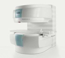
From Siemens Medical Systems;
A new, powerful, compact player in MRI. For both, patients and health care professionals, the mid-field has realized a giant step to cost efficient quality care. Obese patients and people with claustrophobia appreciate the comfortable side loading. The smallest pole diameter - 137 cm (54 inches) allows for optimal patient comfort.
Device Information and Specification
CLINICAL APPLICATION
Whole body
SE, FLASH, FISP, IR, FIR, STIR, TrueIR/FISP, FSE, MT, SS-FSE, MT-SE, MTC, MSE, EPI, PSIF
IMAGING MODES
Single, multislice, volume study, multi angle, multi oblique
512 x 512 full screen display
41 cm vertical gap distance
| | | |
• View the DATABASE results for 'MAGNETOM C™' (2).
| | | | | | Further Reading: | Basics:
|
|
| |
| | | | | |
| |
|
•
In the 1930's, Isidor Isaac Rabi (Columbia University) succeeded in detecting and measuring single states of rotation of atoms and molecules, and in determining the mechanical and magnetic moments of the nuclei.
•
Felix Bloch (Stanford University) and Edward Purcell (Harvard University) developed instruments, which could measure the magnetic resonance in bulk material such as liquids and solids. (Both honored with the Nobel Prize for Physics in 1952.) [The birth of the NMR spectroscopy]
•
In the early 70's, Raymond Damadian (State University of New York) demonstrated with his NMR device, that there are different T1 relaxation times between normal and abnormal tissues of the same type, as well as between different types of normal tissues.
•
In 1973, Paul Lauterbur (State University of New York) described a new imaging technique that he termed Zeugmatography. By utilizing gradients in the magnetic field, this technique was able to produce a two-dimensional image (back-projection). (Through analysis of the characteristics of the emitted radio waves, their origin could be determined.) Peter Mansfield further developed the utilization of gradients in the magnetic field and the mathematically analysis of these signals for a more useful imaging technique. (Paul C Lauterbur and Peter Mansfield were awarded with the 2003 Nobel Prize in Medicine.)
•
1977/78: First images could be presented.
A cross section through a finger by Peter Mansfield and Andrew A. Maudsley.
Peter Mansfield also could present the first image through the abdomen.
•
In 1977, Raymond Damadian completed (after 7 years) the first MR scanner (Indomitable). In 1978, he founded the FONAR Corporation, which manufactured the first commercial MRI scanner in 1980. Fonar went public in 1981.
•
1981: Schering submitted a patent application for Gd-DTPA dimeglumine.
•
1982: The first 'magnetization-transfer' imaging by Robert N. Muller.
•
In 1983, Toshiba obtained approval from the Ministry of Health and Welfare in Japan for the first commercial MRI system.
•
1986: Jürgen Hennig, A. Nauerth, and Hartmut Friedburg (University of Freiburg) introduced RARE (rapid acquisition with relaxation enhancement) imaging. Axel Haase, Jens Frahm, Dieter Matthaei, Wolfgang Haenicke, and Dietmar K. Merboldt (Max-Planck-Institute, Göttingen) developed the FLASH ( fast low angle shot) sequence.
•
1988: Schering's MAGNEVIST gets its first approval by the FDA.
•
In 1991, fMRI was developed independently by the University of Minnesota's Center for Magnetic Resonance Research (CMRR) and Massachusetts General Hospital's (MGH) MR Center.
•
From 1992 to 1997 Fonar was paid for the infringement of it's patents from 'nearly every one of its competitors in the MRI industry including giant multi-nationals as Toshiba, Siemens, Shimadzu, Philips and GE'.
| | | | | |
• View the DATABASE results for 'MRI History' (6).
| | |
• View the NEWS results for 'MRI History' (1).
| | | | | | Further Reading: | | Basics:
|
|
News & More:
| |
| |
| | | Searchterm 'FLASH' was also found in the following services: | | | | |
| | |
| |
|
The subacute risks and side effects of magnetic and RF fields (for patients and staff) have been intensively examined for a long time, but there have been no long-term studies following persons who have been exposed to the static magnetic fields used in MRI. However, no permanent hazardous effects of a static magnetic field exposure upon human beings have yet been demonstrated.
Temporary possible side effects of high magnetic and RF fields:
•
Varying magnetic fields can induce so-called magnetic phosphenes that occur when an individual is subject to rapid changes of 2-5 T/s, which can produce a flashing sensation in the eyes. This temporary side effect does not seem to damage the eyes. Static field strengths used for clinical MRI examinations vary between 0.2 and 3.0 tesla;; field changes during the MRI scan vary in the dimension of mT/s. Experimental imaging units can use higher field strengths of up to 14.0 T, which are not approved for human use.
•
The Radio frequency pulses mainly produce heat, which is absorbed by the body tissue. If the power of the RF radiation is very high, the patient may be heated too much. To avoid this heating, the limit of RF exposure in MRI is up to the maximum specific absorption rate (SAR) of 4 W/kg whole body weight (can be different from country to country). For MRI safety reasons, the MRI machine starts no sequence, if the SAR limit is exceeded.
•
Very high static magnetic fields are needed to reduce the conductivity of nerves perceptibly. Augmentation of T waves is observed at fields used in standard imaging but this side effect in MRI is completely reversible upon removal from the magnet. Cardiac arrhythmia threshold is typically set to 7-10 tesla. The magnetohydrodynamic effect, which results from a voltage occurring across a vessel in a magnetic field and percolated by a saline solution such as blood, is irrelevant at the field strengths used.
The results of some animal and cellular studies suggest the possibility that electromagnetic fields may act as co-carcinogens or tumor promoters, but the data are inconclusive.
Up to 45 tesla, no important effects on enzyme systems have been observed. Neither changes in enzyme kinetics, nor orientation changes in macromolecules have been conclusively demonstrated.
There are some publications associating an increase in the incidence of leukemia with the location of buildings close to high-current power lines with extremely low-frequency (ELF) electromagnetic radiation of 50-60 Hz, and industrial exposure to electric and magnetic fields but a transposition of such effects to MRI or MRS seems unlikely.
Under consideration of the MRI safety guidelines, real dangers or risks of an exposure with common MRI field strengths up to 3 tesla as well as the RF exposure during the MRI scan, are not to be expected.
For more MRI safety information see also Nerve Conductivity,
Contraindications, Pregnancy
and Specific Absorption Rate.
See also the related poll result: ' In 2010 your scanner will probably work with a field strength of' | | | |
• View the DATABASE results for 'MRI Risks' (9).
| | |
• View the NEWS results for 'MRI Risks' (3).
| | | | | | Further Reading: | Basics:
|
|
News & More:
| |
| |
| | | | | | | | | |
| | | |
|
| |
 | Look
Ups |
| |


