Magnetic Resonance - Technology Information Portal Welcome to MRI Technology
Info
  Sheets

Out-
      side


 'Blurring' 
SEARCH FOR    
 
  2 3 5 A B C D E F G H I J K L M N O P Q R S T U V W X Y Z
Result : Searchterm 'Blurring' found in 1 term [] and 14 definitions []
previous     6 - 10 (of 15)     next
Result Pages : [1]  [2 3]
Searchterm 'Blurring' was also found in the following services: 
spacer
Forum  (6)  
 
Blood Oxygenation Level Dependent ContrastInfoSheet: - Sequences - 
Intro, 
Overview, 
Types of, 
etc.MRI Resource Directory:
 - Functional MRI -
 
(BOLD) In MRI the changes in blood oxygenation level are visible. Oxyhaemoglobin (the principal haemoglobin in arterial blood) has no substantial magnetic properties, but deoxyhaemoglobin (present in the draining veins after the oxygen has been unloaded in the tissues) is strongly paramagnetic. It can thus serve as an intrinsic paramagnetic contrast agent in appropriately performed brain MRI. The concentration and relaxation properties of deoxyhaemoglobin make it a susceptibility , e.g. T2 relaxation effective contrast agent with little effect on T1 relaxation.
During activation of the brain, the oxygen consumption of the local tissue increase by approximately 5% with that the oxygen tension will decrease. As a consequence, after a short period of time vasodilatation occurs, resulting in a local increase of blood volume and flow by 20 - 40%. The incommensurate change in local blood flow and oxygen extraction increases the local oxygen level.
By using T2 weighted gradient echo EPI sequences, which are highly susceptibility sensitive and fast enough to capture the three-dimensional nature of activated brain areas will show an increase in signal intensity as oxyhaemoglobin is diamagnetic and deoxyhaemoglobin is paramagnetic. Other MR pulse sequences, such as spoiled gradient echo pulse sequences are also used.
As the effects are subtle and of the order of 2% in 1.5 T MR imaging, sophisticated methodology, paradigms and data analysis techniques have to be used to consistently demonstrate the effect.
As the BOLD effect is due to the deoxygenated blood in the draining veins, the spatial localization of the region where there is increased blood flow resulting in decreased oxygen extraction is not as precisely defined as the morphological features in MRI. Rather there is a physiological blurring, and is estimated that the linear dimensions of the physiological spatial resolution of the BOLD phenomenon are around 3 mm at best.
spacer
 
• Related Searches:
    • Oxygen Mapping
    • Susceptibility
    • Blood Flow Imaging
    • T2 Relaxation
    • Perfusion Imaging
 
Further Reading:
  Basics:
IMAGE CONTRAST IN MRI(.pdf)
   by www.assaftal.com    
Vascular Filters of Functional MRI: Spatial Localization Using BOLD and CBV Contrast
  News & More:
A mechanistic computational framework to investigate the hemodynamic fingerprint of the blood oxygenation level-dependent signal
Tuesday, 29 August 2023   by analyticalsciencejournals.onlinelibrary.wiley.com    
The utility of texture analysis of kidney MRI for evaluating renal dysfunction with multiclass classification model
Tuesday, 30 August 2022   by www.nature.com    
MRI Technique Used to Identify Future Risk of Binge Drinking
Monday, 6 January 2020   by www.diagnosticimaging.com    
Gold Acupuncture Needle MRI Pain Discovery
Friday, 3 January 2014   by www.healthcmi.com    
MRI method for measuring MS progression validated
Thursday, 19 December 2013   by www.eurekalert.org    
Searchterm 'Blurring' was also found in the following service: 
spacer
Radiology  (4) Open this link in a new window
Cardiac Gating
 
This method synchronize the heartbeat with the beginning of the TR, whereat the r wave is used as the trigger. Cardiac gating times the acquisition of MR data to physiological motion in order to minimize motion artifacts. ECG gating techniques are useful whenever data acquisition is too slow to occur during a short fraction of the cardiac cycle.
Image blurring due to cardiac-induced motion occurs for imaging times of above approximately 50 ms in systole, while for imaging during diastole the critical time is of the order of 200-300 ms. The acquisition of an entire image in this time is only possible with using ultrafast MR imaging techniques. If a series of images using cardiac gating or real-time echo planar imaging EPI are acquired over the entire cardiac cycle, pixel-wise velocity and vascular flow can be obtained.
In simple cardiac gating, a single image line is acquired in each cardiac cycle. Lines for multiple images can then be acquired successively in consecutive gate intervals. By using the standard multiple slice imaging and a spin echo pulse sequence, a number of slices at different anatomical levels is obtained. The repetition time (TR) during a ECG-gated acquisition equals the RR interval, and the RR interval defines the minimum possible repetition time (TR). If longer TRs are required, multiple integers of the RR interval can be selected. When using a gradient echo pulse sequence, multiple phases of a single anatomical level or multiple slices at different anatomical levels can be acquired over the cardiac cycle.
Also called cardiac triggering.
 
Images, Movies, Sliders:
 Cardiac Infarct Short Axis Cine Overview  Open this link in a new window
    

Courtesy of  Robert R. Edelman
 Infarct 4 Chamber Cine  Open this link in a new window
    

Courtesy of  Robert R. Edelman
 
spacer

• View the DATABASE results for 'Cardiac Gating' (15).Open this link in a new window

 
Further Reading:
  Basics:
Cardiac MRI - Technical Aspects Primer
Wednesday, 7 August 2002
Electrocardiogram in an MRI Environment: Clinical Needs, Practical Considerations, Safety Implications, Technical Solutions and Future Directions
Wednesday, 25 January 2012   by cdn.intechopen.com    
Motion-compensation of Cardiac Perfusion MRI using a Statistical Texture Ensemble(.pdf)
June 2003   by www.imm.dtu.dk    
MRI Resources 
MR Guided Interventions - Homepages - Devices - Colonography - MRI Physics - Service and Support
 
Double Inversion Recovery T1 MeasurementInfoSheet: - Sequences - 
Intro, 
Overview, 
Types of, 
etc.MRI Resource Directory:
 - Sequences -
 
(DIR or DIRT1) Double inversion recovery T1 measurement is a T1 weighted black blood MRA sequence in which the signal from blood is suppressed. The inversion time to suppress blood is described as the duration between the initial inversion pulse and time point that the longitudinal magnetization of blood reaches the zero point. The readout starts at the blood suppression inversion time (BSP TI) and blood in the imaging slice gives no signal. This inversion time is around 650 ms with a 60 beat per minute heart rate at 1.5 T.
The TI can be decreased by using a wider receive bandwidth, shorter echo train length and/or narrow trigger window. Wide bandwidth also decreases the blurring caused by long echo trains at the expense of signal to noise ratio. In case of in plane or slow flow the suppression of the signal from blood may be incomplete. With increased TE or change of the image plane the blood suppression can be improved.
Double inversion recovery is a breath hold technique with one image per acquisition used in cardiovascular imaging. The patient is instructed to hold the breath in expiration (if not possible also inspiration can be taken), so that the end diastolic volume in the cardiac chambers would be the same during entire scanning. DIR provides fine details of the boundary between the lumen and the wall of the cardiac chambers and main vascular and heart structures, pericardium, and mediastinal tissues.
 
Images, Movies, Sliders:
 Normal Dual Inversion Fast Spin-echo  Open this link in a new window
      

Courtesy of  Robert R. Edelman

 
spacer

• View the DATABASE results for 'Double Inversion Recovery T1 Measurement' (2).Open this link in a new window

 
Further Reading:
  News & More:
Artificial double inversion recovery images can substitute conventionally acquired images: an MRI-histology study
Wednesday, 16 February 2022   by www.nature.com    
Searchterm 'Blurring' was also found in the following services: 
spacer
Forum  (6)  
 
Fast Spin EchoForum -
related threadsInfoSheet: - Sequences - 
Intro, 
Overview, 
Types of, 
etc.
 
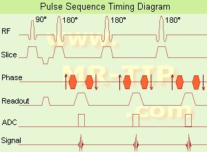
Fast Spin Echo Diagram (FSE) In the pulse sequence timing diagram, a fast spin echo sequence with an echo train length of 3 is illustrated. This sequence is characterized by a series of rapidly applied 180° rephasing pulses and multiple echoes, changing the phase encoding gradient for each echo.
The echo time TE may vary from echo to echo in the echo train. The echoes in the center of the K-space (in the case of linear k-space acquisition) mainly produce the type of image contrast, whereas the periphery of K-space determines the spatial resolution. For example, in the middle of K-space the late echoes of T2 weighted images are encoded. T1 or PD contrast is produced from the early echoes.
The benefit of this technique is that the scan duration with, e.g. a turbo spin echo turbo factor / echo train length of 9, is one ninth of the time. In T1 weighted and proton density weighted sequences, there is a limit to how large the ETL can be (e.g. a usual ETL for T1 weighted images is between 3 and 7). The use of large echo train lengths with short TE results in blurring and loss of contrast. For this reason, T2 weighted imaging profits most from this technique.
In T2 weighted FSE images, both water and fat are hyperintense. This is because the succession of 180° RF pulses reduces the spin spin interactions in fat and increases its T2 decay time. Fast spin echo (FSE) sequences have replaced conventional T2 weighted spin echo sequences for most clinical applications. Fast spin echo allows reduced acquisition times and enables T2 weighted breath hold imaging, e.g. for applications in the upper abdomen.
In case of the acquisition of 2 echoes this type of a sequence is named double fast spin echo / dual echo sequence, the first echo is usually density and the second echo is T2 weighted image. Fast spin echo images are more T2 weighted, which makes it difficult to obtain true proton density weighted images. For dual echo imaging with density weighting, the TR should be kept between 2000 - 2400 msec with a short ETL (e.g., 4).
Other terms for this technique are:
Turbo Spin Echo
Rapid Imaging Spin Echo,
Rapid Spin Echo,
Rapid Acquisition Spin Echo,
Rapid Acquisition with Refocused Echoes
 
Images, Movies, Sliders:
 Lumbar Spine T2 FSE Sagittal  Open this link in a new window
    

Courtesy of  Robert R. Edelman
 MRI - Anatomic Imaging of the Foot  Open this link in a new window
    
SlidersSliders Overview

 Lumbar Spine T2 FSE Axial  Open this link in a new window
    

Courtesy of  Robert R. Edelman
 
spacer

• View the DATABASE results for 'Fast Spin Echo' (31).Open this link in a new window

 
Further Reading:
  Basics:
MYELIN-SELECTIVE MRI: PULSE SEQUENCE DESIGN AND OPTIMIZATION
   by www.imaging.robarts.ca    
Advances in Magnetic Resonance Neuroimaging
Friday, 27 February 2009   by www.ncbi.nlm.nih.gov    
  News & More:
New MR sequence helps radiologists more accurately evaluate abnormalities of the uterus and ovaries
Thursday, 23 April 2009   by www.eurekalert.org    
Spin echoes, CPMG and T2 relaxation - Introductory NMR & MRI from Magritek
2013   by www.azom.com    
Searchterm 'Blurring' was also found in the following service: 
spacer
Radiology  (4) Open this link in a new window
Fast Spoiled Grass Sequence with IR PreparationInfoSheet: - Sequences - 
Intro, 
Overview, 
Types of, 
etc.
 
(IR-FSPGR) This sequence uses an inversion pulse followed by a spoiled low flip angle and short TR gradient echo train to collect all partition data. The 3D-IR-FSPGR uses centric ordering of Gz. The selected TI is also the effective TI. The centric reordering strategy can lead to image blurring if a steady state is not achieved quickly with low flip angles (less than 20°).
spacer
MRI Resources 
Service and Support - Mobile MRI - Breast MRI - MRI Centers - MRI Accidents - Contrast Agents
 
previous      6 - 10 (of 15)     next
Result Pages : [1]  [2 3]
 Random Page
 
Share This Page
FacebookTwitterLinkedIn

MR-TIP    
Community   
User
Pass
Forgot your UserID/Password ?    


New acceleration techniques will :
reduce scan times 
cause artifacts 
increase expenses 
be useful if you have a lot of experience 
doesn't do much 
never heard of 

Look
      Ups


MR-TIP.com uses cookies! By browsing MR-TIP.com, you agree to our use of cookies.

Magnetic Resonance - Technology Information Portal
Member of SoftWays' Medical Imaging Group - MR-TIP • Radiology-TIP • Medical-Ultrasound-Imaging • 
Copyright © 2003 - 2024 SoftWays. All rights reserved. [ 28 April 2024]
Terms of Use | Privacy Policy | Advertising
 [last update: 2024-02-26 03:41:00]