Magnetic Resonance - Technology Information Portal Welcome to MRI Technology
Info
  Sheets

Out-
      side


 'Array' 
SEARCH FOR    
 
  2 3 5 A B C D E F G H I J K L M N O P Q R S T U V W X Y Z
Result : Searchterm 'Array' found in 8 terms [] and 53 definitions []
previous     21 - 25 (of 61)     next
Result Pages : [1 2]  [3 4 5 6 7 8 9 10 11 12 13]
Searchterm 'Array' was also found in the following services: 
spacer
News  (5)  Resources  (4)  Forum  (5)  
 
Opera (E-SCAN™ XQ)InfoSheet: - Devices -
Intro, 
Types of Magnets, 
Overview, 
etc.MRI Resource Directory:
 - Devices -
 
www.esaote.com/products/MRI/eScanXQ/products1.htm Manufactured by Esaote S.p.A.;
a low field open MRI scanner with permanent magnet for orthopedic use. The outstanding feature of this MRI system is a patient friendly design with 24 cm diameter, which allows the imaging of extremities and small body parts like shoulder MRI. The power consumption is around 1.3 kW and the needed minimum floor space is an area of 16 sq m.
At RSNA 2006 Hologic Inc. introduced a new dedicated extremity MRI scanner, the Opera. Manufactured by Esaote is the Opera a redesign of Esaote's 0.2 Tesla E-Scan XQ platform, which now enables complete imaging of all extremities, including hip and shoulder applications. 'Real-time positioning' reportedly speeds patient setup and reduces exam times.
Esaote North America and Hologic Inc are the U.S. distributors of this MRI device.
Device Information and Specification
CLINICAL APPLICATION
Dedicated extremity
CONFIGURATION
Extremity, shoulder (2), flex coil, knee dual phased array, ankle//foot dual phased array, hand//wrist dual phased array coil
PULSE SEQUENCES
SE, GE, IR, STIR, FSE, 3D CE, GE-STIR, 3D GE, ME, TME, HSE
IMAGING MODES
Single, multislice, volume study, fast scan, multi slab
TR
10 - 5000 msec
TE
6 - 110 msec
SINGLE SLICE
0.6 sec/image
MULTISLICE
0.6 sec/image
17 cm
2D: 2 mm - 10 mm;
3D: 0.6 mm - 10 mm
MEASURING MATRIX
256 x 256 maximum
PIXEL INTENSITY
4096 gray lvls, 256 lvls in 3D
MAGNET TYPE
Permanent
24 cm H, open
MAGNET WEIGHT
2250 kg, 4960 lbs
H*W*D
79 x 65 x 85 cm
POWER REQUIREMENTS
2,0 kW; 110/220 V single phase
STRENGTH
20 mT/m
5-GAUSS FRINGE FIELD, radial/axial
150 cm/130 cm
Passive
spacer
 
Further Reading:
  News & More:
E-Scan, 510(k) Summary(.pdf)
Saturday, 15 May 2004   by www.accessdata.fda.gov    
MRI Resources 
Safety pool - Devices - Fluorescence - MRI Training Courses - Service and Support - Bioinformatics
 
Parallel Imaging TechniqueForum -
related threadsInfoSheet: - Sequences - 
Intro, 
Overview, 
Types of, 
etc.
 
In parallel MR imaging, a reduced data set in the phase encoding direction(s) of k-space is acquired to shorten acquisition time, combining the signal of several coil arrays. The spatial information related to the phased array coil elements is utilized for reducing the amount of conventional Fourier encoding.
First, low-resolution, fully Fourier-encoded reference images are required for sensitivity assessment. Parallel imaging reconstruction in the Cartesian case is efficiently performed by creating one aliased image for each array element using discrete Fourier transformation. The next step then is to create an full FOV image from the set of intermediate images. Parallel reconstruction techniques can be used to improve the image quality with increased signal to noise ratio, spatial resolution, reduced artifacts, and the temporal resolution in dynamic MRI scans.
Parallel imaging algorithms can be divided into 2 main groups:
Image reconstruction produced by each coil (reconstruction in the image domain, after Fourier transform): SENSE (Sensitivity Encoding), PILS (Partially Parallel Imaging with Localized Sensitivity), ASSET.
Reconstruction of the Fourier plane of images from the frequency signals of each coil (reconstruction in the frequency domain, before Fourier transform): GRAPPA.
Additional techniques include SMASH, SPEEDER™, IPAT (Integrated Parallel Acquisition Techniques - derived of GRAPPA a k-space based technique) and mSENSE (an image based enhanced version of SENSE).
 
Images, Movies, Sliders:
 Circle of Willis, Time of Flight, MIP  Open this link in a new window
    
SlidersSliders Overview

 
spacer

• View the DATABASE results for 'Parallel Imaging Technique' (12).Open this link in a new window

 
Further Reading:
  Basics:
Parallel MRI Using Multiple Receiver Coils
   by www-math.mit.edu    
Coil Arrays for Parallel MRI: Introduction and Overview.
   by www.mr.ethz.ch    
  News & More:
Cardiac MRI Becoming More Widely Available Thanks to AI and Reduced Exam Times
Wednesday, 19 February 2020   by www.dicardiology.com    
The Effects of Breathing Motion on DCE-MRI Images: Phantom Studies Simulating Respiratory Motion to Compare CAIPIRINHA-VIBE, Radial-VIBE, and Conventional VIBE
Tuesday, 7 February 2017   by www.kjronline.org    
Implementation of Dual-Source RF Excitation in 3 T MR-Scanners Allows for Nearly Identical ADC Values Compared to 1.5 T MR Scanners in the Abdomen
Wednesday, 29 February 2012   by www.plosone.org    
Clinical evaluation of a speed optimized T2 weighted fast spin echo sequence at 3.0 T using variable flip angle refocusing, half-Fourier acquisition and parallel imaging
Wednesday, 25 October 2006
MRI Resources 
Open Directory Project - PACS - Process Analysis - Most Wanted - Safety Products - Mass Spectrometry
 
Sense CoilInfoSheet: - Coils - 
Intro, 
Overview, 
etc.MRI Resource Directory:
 - Coils -
 
An RF array coil consisting of 2 - 6 several separately resonant elements, 2 or more of which can be selected as the receiver coil at a particular time. Coils not in use are decoupled. The spatial information related to the coils of a receiver array are utilized, reference images are required, obtained with each array element and with a body coil. Fast parallel imaging techniques using surface multicoils, such as sensitivity encoding (SENSE) or simultaneous acquisition of spatial harmonics (SMASH), have recently raised interest in the field of MR imaging.

See also the related poll result: '3rd party coils are better than the original manufacturer coils'
spacer

• View the DATABASE results for 'Sense Coil' (2).Open this link in a new window

 
Further Reading:
  Basics:
System Architecture
2003   by www.revisemri.com    
Searchterm 'Array' was also found in the following services: 
spacer
News  (5)  Resources  (4)  Forum  (5)  
 
Imaging CoilInfoSheet: - Coils - 
Intro, 
Overview, 
etc.MRI Resource Directory:
 - Coils -
 
Imaging coils are radio frequency coils used in magnetic resonance imaging for sending and/or receiving electromagnetic radiation. Several MR imaging coils in different shapes and sizes are necessary for different body parts and to handle individual applications.
For Example:
Birdcage coils provide high homogeneity and good signal to noise ratio (SNR) in brain MRI scans.
Surface coils with small coil diameter and higher SNR enable to image the temporomandibular joints (TMJ).
The implemented body coil allows the scanning of body parts with large field of views.
Micro coils are available to image finger joints.
High performance phased array coils are today state-of-the-art for a wide range of applications from head, spine, knee or shoulder to cardiac MRI.
For different types of coils see Volume Coil, Sense Coil, Array Coil, Surface Coil, and Bird Cage Coil.

See also the related poll result: '3rd party coils are better than the original manufacturer coils'
spacer

• View the DATABASE results for 'Imaging Coil' (7).Open this link in a new window


• View the NEWS results for 'Imaging Coil' (9).Open this link in a new window.
 
Further Reading:
  Basics:
Radio-frequency Coil Selection for MR Imaging of the Brain and Skull Base1
   by radiology.rsnajnls.org    
MRI Resources 
Pathology - MRI Technician and Technologist Jobs - Distributors - Case Studies - Education pool - Sequences
 
MAGNETOM C™InfoSheet: - Devices -
Intro, 
Types of Magnets, 
Overview, 
etc.MRI Resource Directory:
 - Devices -
 
www.medical.siemens.com 
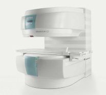
From Siemens Medical Systems;
A new, powerful, compact player in MRI. For both, patients and health care professionals, the mid-field has realized a giant step to cost efficient quality care. Obese patients and people with claustrophobia appreciate the comfortable side loading. The smallest pole diameter - 137 cm (54 inches) allows for optimal patient comfort.
Device Information and Specification
CLINICAL APPLICATION
Whole body
CONFIGURATION
C-shaped open MRI
Multi channel imaging, CP Head//Neck Array Coil, Body/Spine Array Coil, large, Transmit Coil
SYNCHRONIZATION
ECG/peripheral: Optional/yes, respiratory gating
PULSE SEQUENCES
SE, FLASH, FISP, IR, FIR, STIR, TrueIR/FISP, FSE, MT, SS-FSE, MT-SE, MTC, MSE, EPI, PSIF
IMAGING MODES
Single, multislice, volume study, multi angle, multi oblique
TR
Min. TR 2.81 ms
TE
Min. TE 0.98 ms
512 x 512 full screen display
MEASURING MATRIX
64 x 64 to 512 x 512
FOV
0.5 - 40 cm
MAGNET TYPE
Permanent
BORE DIAMETER
or W x H
41 cm vertical gap distance
MAGNET WEIGHT
16000 kg
H*W*D
233 x 206 x 160 cm
STRENGTH
24 mT/m
5-GAUSS FRINGE FIELD
2.2 m / 2.2 m
Passive and active
spacer

• View the DATABASE results for 'MAGNETOM C™' (2).Open this link in a new window

 
Further Reading:
  Basics:
Section 2: 510(k) Summary, MAGNETOM C! System Classification Name: Magnetic Resonance Diagnostic(.pdf)
   by www.accessdata.fda.gov    
MRI Resources 
Anatomy - MRI Physics - Mobile MRI Rental - Research Labs - Universities - Diffusion Weighted Imaging
 
previous      21 - 25 (of 61)     next
Result Pages : [1 2]  [3 4 5 6 7 8 9 10 11 12 13]
 Random Page
 
Share This Page
FacebookTwitterLinkedIn

MR-TIP    
Community   
User
Pass
Forgot your UserID/Password ?    


MRI is trending to low field magnets :
reduced costs will lead to this change 
AI will close the gap to high field 
only in remote areas 
is only temporary 
never 

Look
      Ups


MR-TIP.com uses cookies! By browsing MR-TIP.com, you agree to our use of cookies.

Magnetic Resonance - Technology Information Portal
Member of SoftWays' Medical Imaging Group - MR-TIP • Radiology-TIP • Medical-Ultrasound-Imaging • 
Copyright © 2003 - 2024 SoftWays. All rights reserved. [ 4 May 2024]
Terms of Use | Privacy Policy | Advertising
 [last update: 2024-02-26 03:41:00]