 | Info
Sheets |
| | | | | | | | | | | | | | | | | | | | | | | | |
 | Out-
side |
| | | | |
|
| | | | |
Result : Searchterm 'tensor' found in 2 terms [ ] and 14 definitions [ ] and 14 definitions [ ] ]
| | previous 11 - 15 (of 16) nextResult Pages : [1] [2 3 4] |  | |  | Searchterm 'tensor' was also found in the following services: | | | | |
| |  |
| |
|
Rapid echo planar imaging and high-performance MRI gradient systems create fast-switching magnetic fields that can stimulate muscle and nerve tissues produced by either changing the electrical resistance or the potential of the excitation. There are apparently no effects on the conduction of impulses in the nerve fiber up to field strength of 0.1 T. A preliminary study has indicated neurological effects by exposition to a whole body imager at 4.0 T. Theoretical examinations argue that field strengths of 24 T are required to produce a 10% reduction of nerve impulse conduction velocity.
Nerve stimulations during MRI scans can be induced by very rapid changes of the magnetic field. This stimulation may occur for example during diffusion weighted sequences or diffusion tensor imaging and can result in muscle contractions caused by effecting motor nerves. The so-called magnetic phosphenes are attributed to magnetic field variations and may occur in a threshold field change of between 2 and 5 T/s. Phosphenes are stimulations of the optic nerve or the retina, producing a flashing light sensation in the eyes. They seem not to cause any damage in the eye or the nerve.
Varying magnetic fields are also used to stimulate bone-healing in non-unions and pseudarthroses. The reasons why pulsed magnetic fields support bone-healing are not completely understood. The mean threshold levels for various stimulations are 3600 T/s for the heart, 900 T/s for the respiratory system, and 60 T/s for the peripheral nerves.
Guidelines in the United States limit switching rates at a factor of three below the mean threshold for peripheral nerve stimulation. In the event that changes in nerve conductivity happens, the MRI scan parameters should be adjusted to reduce dB/dt for nerve stimulation. | | | | • For this and other aspects of MRI safety see our InfoSheet about MRI Safety. | | | • Patient-related information is collected in our MRI Patient Information.
| | | | |  Further Reading: Further Reading: | | Basics:
|
|
News & More:
| |
| |
| |  | MRI Safety Resources | | | | |
| | | |
| |
|
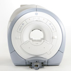
From GE Healthcare;
The GE Signa HDx MRI system is a whole body magnetic resonance scanner designed to support high resolution, high signal to noise ratio, and short scan times.
The 1.5T Signa HDx MR Systems is a modification of the currently marketed GE 1.5T machines, with the main difference being the change to the receive chain architecture that includes a thirty two independent receive channels, and allows for future expansion in 16 channel increments. The overall system has been improved with a simplified user interface
and a single 23" liquid crystal display, improved multi channel surface coil connectivity, and an improved image reconstruction architecture known as the Volume Recon Engine (VRE).
Device Information and Specification CLINICAL APPLICATION Whole body CONFIGURATION Compact short bore Standard: SE, IR, 2D/3D GRE and SPGR, Angiography: 2D/3D TOF, 2D/3D Phase Contrast; 2D/3D FSE, 2D/3D FGRE and FSPGR, SSFP, FLAIR, EPI, optional: 2D/3D Fiesta, FGRET, Spiral, Tensor, 2D 0.7 mm to 20 mm; 3D 0.1 mm to 5 mm 128x512 steps 32 phase encode POWER REQUIREMENTS 480 or 380/415 less than 0.03 L/hr liquid helium | | | | | |
| | | | | |
| |
|

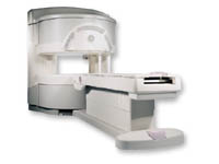
From GE Healthcare;
three dedicated MRI systems - a neuro imager, a cardiovascular system, and a whole body scanner - all in one system.
Device Information and Specification CLINICAL APPLICATION Whole body Head and body coil standard; all other coils optional; open architecture makes system compatible with a wide selection of coils Optional 2D/3D brain and prostate Standard: SE, IR, 2D/3D GRE and SPGR, Angiography: 2D/3D TOF, 2D/3D Phase Contrast;; 2D/3D FSE, 2D/3D FGRE and FSPGR, SSFP, FLAIR, EPI, optional: 2D/3D Fiesta, FGRET, Spiral, TensorTR 1.2 to 12000 msec in increments of 1 msec TE 0.4 to 2000 msec in increments of 1 msec 2D 0.7 mm to 20 mm; 3D 0.1 mm to 5 mm 128x512 steps 32 phase encode POWER REQUIREMENTS 480 or 380/415 Less than 0.03 L/hr liquid He STRENGTH Zoom 40 mT/m, whole body 23 mT/m 4.0 m x 2.8 m axial x radial | | | |
• View the DATABASE results for 'Signa Infinity 1.5T™ TwinSpeed with Excite' (2).
| | | | | | Further Reading: | News & More:
|
|
| |
| | | Searchterm 'tensor' was also found in the following services: | | | | |
| | |
| |
|

From GE Healthcare;
'EXCITE technology has the potential to open the door to new imaging techniques and clinical applications, leaping beyond conventional two and three-dimensional MRI to true 4D imaging that will improve the diagnosis of disease throughout the human body from head to foot.' Robert R. Edelman, M.D., Professor of Radiology at Northwestern University Medical School and Chairman, Department of Radiology, at Evanston Northwestern Healthcare.
Device Information and Specification CLINICAL APPLICATION Whole body Head and body coil standard; all other coils optional; open architecture makes system compatible with a wide selection of coils Optional 2D/3D brain and prostate Standard: SE, IR, 2D/3D GRE and SPGR, Angiography: 2D/3D TOF, 2D/3D Phase Contrast;; 2D/3D FSE, 2D/3D FGRE and FSPGR, SSFP, FLAIR, EPI, optional: 2D/3D Fiesta, FGRET, Spiral, TensorTR 1.3 to 12000 msec in increments of 1 msec TE 0.4 to 2000 msec in increments of 1 msec 2D 0.7 mm to 20 mm; 3D 0.1 mm to 5 mm 128x512 steps 32 phase encode 0.08 mm; 0.02 mm optional POWER REQUIREMENTS 480 or 380/415 less than 0.03 L/hr liquid heliumSTRENGTH SmartSpeed 23 mT/m, HiSpeed Plus 33 mT/m, EchoSpeed Plus 33 mT/m 4.0 m x 2.8 m axial x radial | | | |
• View the DATABASE results for 'Signa Infinity 1.5T™ with Excite' (2).
| | | | |
| | | | | |
| |
|
From GE Healthcare;
a friendly and less confining appearance targets the 7% of individuals who refuse to have an MRI because of claustrophobia. This open MRI system is also up to three times faster than other scanners, therefore the Signa OpenSpeed™ reducing exam time and scheduling
issues. In addition, a swing table provides better access and supports up to 500 pounds.
Device Information and Specification CLINICAL APPLICATION Whole body Standard: SE, IR, 2D/3D GRE and SPGR, Angiography: 2D/3D TOF, 2D/3D Phase Contrast;; 2D/3D FSE, 2D/3D FGRE and FSPGR, SSFP, FLAIR, EPI, optional: 2D/3D Fiesta, FGRET, Spiral, TensorTR 1.3 to 12000 msec in increments of 1 msec TE 0.4 to 2000 msec in increments of 1 msec 2D: 0.8mm - 20mm 3D: 0.1mm - 20mm 0.08 mm; 0.02 mm optional POWER REQUIREMENTS 200 - 480, 3-phase | | | |
• View the DATABASE results for 'Signa OpenSpeed™' (2).
| | | | | | Further Reading: | News & More:
|
|
| |
| | | | |
| | | |
|
| |
 | Look
Ups |
| |


