Magnetic Resonance - Technology Information Portal Welcome to MRI Technology
Info
  Sheets

Out-
      side


 'temporal' 
SEARCH FOR    
 
  2 3 5 A B C D E F G H I J K L M N O P Q R S T U V W X Y Z
Result : Searchterm 'temporal' found in 1 term [] and 25 definitions []
previous     21 - 25 (of 26)     next
Result Pages : [1]  [2 3 4 5 6]
Searchterm 'temporal' was also found in the following services: 
spacer
News  (8)  Forum  (1)  
 
Segmented K-Space Data Acquisition
 
A set of k-space lines collected in a specified order but not constituting a complete coverage of k-space, thus can be used in conjunction with all ultrafast MRI techniques. Several segmental acquisitions may need to be run for complete coverage of k-space. If these lines are recorded for a single rather than multiple images, imaging time can be shortened considerably maintaining an acceptable temporal resolution.
For example, rapidly acquiring eight k-space lines per segment after each trigger until 128 lines of k-space are acquired in 16 triggers, thus makes image acquisition of multiple cardiac phases or anatomical slices possible in a breath-hold.
spacer
MRI Resources 
Online Books - PACS - Fluorescence - Blood Flow Imaging - Equipment - Abdominal Imaging
 
Simultaneous Acquisition of Spatial Harmonics
 
(SMASH) Several lines of data are acquired for each phase encoding step, which is also referred to as a k-space trajectory.
SMASH imaging with a four-element array coil is four times faster and can be used to achieve almost real-time imaging. The maximum reduction in acquisition time is determined by the number of array coil elements. Thus, the heart can be scanned with higher temporal resolution and increased spatial resolution.
SMASH and SENSE differ from other techniques in which only one line of k-space data is acquired for each phase encoding gradient step.

See Sensitivity encoding.
spacer

• View the DATABASE results for 'Simultaneous Acquisition of Spatial Harmonics' (3).Open this link in a new window

MRI Resources 
Implant and Prosthesis - Breast MRI - Breast Implant - Musculoskeletal and Joint MRI - Non-English - Pathology
 
Special ImagingInfoSheet: - Sequences - 
Intro, 
Overview, 
Types of, 
etc.
 
Special imaging primarily means advanced MRI techniques used for qualitative and quantitative measurement of biological metabolism as e.g., spectroscopy, perfusion imaging (PWI, ASL), diffusion weighted imaging (DWI, DTI, DTT) and brain function (BOLD, fMRI). This physiological magnetic resonance techniques offer insights into brain structure, function, and metabolism.
Spectroscopy provides functional information related to identification and quantification of e.g. brain metabolites. MR perfusion imaging has applications in stroke, trauma, and brain neoplasm. MRI provides the high spatial and temporal resolution needed to measure blood flow to the brain. arterial spin labeling techniques utilize the intrinsic protons of blood and brain tissue, labeled by special preparation pulses, rather than exogenous tracers injected into the blood.
MR diffusion tensor imaging characterizes the ability of water to spread across the brain in different directions. Diffusion parallel to nerve fibers has been shown to be greater than diffusion in the perpendicular direction. This provides a tool to study in vivo fiber connectivity in brain MRI.
FMRI allows the detection of a functional activation in the brain because cortical activity is intimately related to local metabolism changes.

See also Diffusion Tensor Tractography.
spacer

• View the NEWS results for 'Special Imaging' (14).Open this link in a new window.
 
Further Reading:
  Basics:
MRI's inside story
Thursday, 4 December 2003   by www.economist.com    
Diffusion Imaging: From Basic Physics to Practical Imaging
1999   by ej.rsna.org    
  News & More:
Philips and University Medical Center Utrecht Partner to Advance Quantitative MRI With MR-STAT
Monday, 10 August 2020   by www.itnonline.com    
This new 'whole body' MRI is better for cancer patients
Tuesday, 28 January 2014   by www.physiciansnews.com    
Turbo-FLASH Based Arterial Spin Labeled Perfusion MRI at 7 T
Thursday, 20 June 2013   by www.plosone.org    
Multiparametric MRI for Detecting Prostate Cancer
Wednesday, 17 December 2014   by www.onclive.com    
Searchterm 'temporal' was also found in the following services: 
spacer
News  (8)  Forum  (1)  
 
Spectral Width
 
The overall width in hertz needed to observe a particular NMR spectrum. This width is generally set using the Nyquist limit; namely, that the temporal sampling rate must be equal to twice the maximum spread in frequencies.
spacer
MRI Resources 
Research Labs - Fluorescence - Chemistry - MR Guided Interventions - Implant and Prosthesis pool - Libraries
 
Ultrafast Gradient Echo SequenceInfoSheet: - Sequences - 
Intro, 
Overview, 
Types of, 
etc.MRI Resource Directory:
 - Sequences -
 
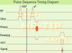
Ultrafast Gradient Echo Sequence Timing Diagram In simple ultrafast GRE imaging, TR and TE are so short, that tissues have a poor imaging signal and - more importantly - poor contrast except when contrast media enhanced (contrast enhanced angiography). Therefore, the magnetization is 'prepared' during the preparation module, most frequently by an initial 180° inversion pulse.
In the pulse sequence timing diagram, the basic ultrafast gradient echo sequence is illustrated. The 180° inversion pulse is executed one time (to the left of the vertical line), the right side represents the data collection period and is often repeated depending on the acquisition parameters.
See also Pulse Sequence Timing Diagram, there you will find a description of the components.
Ultrafast GRE sequences have a short TR,TE, a low flip angle and TR is so short that image acquisition lasts less than 1 second and typically less than 500 ms. Common TR: 3-5 msec, TE: 2 msec, and the flip angle is about 5°. Such sequences are often labeled with the prefix 'Turbo' like TurboFLASH, TurboFFE and TurboGRASS.
This allows one to center the subsequent ultrafast GRE data acquisition around the inversion time TI, where one of the tissues of interest has very little signal as its z-magnetization is passing through zero.
Unlike a standard inversion recovery (IR) sequence, all lines or a substantial segment of k-space image lines are acquired after a single inversion pulse, which can then together be considered as readout module. The readout module may use a variable flip angle approach, or the data acquisition may be divided into multiple segments (shots). The latter is useful particularly in cardiac imaging where acquiring all lines in a single segment may take too long relative to the cardiac cycle to provide adequate temporal resolution.
If multiple lines are acquired after a single pulse, the pulse sequence is a type of gradient echo echo planar imaging (EPI) pulse sequence.

See also Magnetization Prepared Rapid Gradient Echo (MPRAGE) and Turbo Field Echo (TFE).
spacer

• View the DATABASE results for 'Ultrafast Gradient Echo Sequence' (13).Open this link in a new window

MRI Resources 
Stent - Patient Information - Chemistry - Examinations - Breast MRI - Quality Advice
 
previous      21 - 25 (of 26)     next
Result Pages : [1]  [2 3 4 5 6]
 Random Page
 
Share This Page
FacebookTwitterLinkedIn

MR-TIP    
Community   
User
Pass
Forgot your UserID/Password ?    


Personalized protocols (age, gender, body habitus, etc.) lead to :
more automated planning 
improved patient comfort 
better diagnostics 
optimized image quality 
nothing 

Look
      Ups


MR-TIP.com uses cookies! By browsing MR-TIP.com, you agree to our use of cookies.

Magnetic Resonance - Technology Information Portal
Member of SoftWays' Medical Imaging Group - MR-TIP • Radiology-TIP • Medical-Ultrasound-Imaging • 
Copyright © 2003 - 2024 SoftWays. All rights reserved. [ 27 April 2024]
Terms of Use | Privacy Policy | Advertising
 [last update: 2024-02-26 03:41:00]