 | Info
Sheets |
| | | | | | | | | | | | | | | | | | | | | | | | |
 | Out-
side |
| | | | |
|
| | | | | |  | Searchterm 'Meter' was also found in the following services: | | | | |
|  |  |
| |
|
MRI computer can be divided into central processing unit (CPU), consisting of instruction, interpretation and arithmetic unit plus fast access memory, and peripheral devices such as bulk data storage and input and output devices (including, via the interface, the spectrometer).
The computer controls the RF pulses and gradients necessary to acquire data, and process the data to produce spectra or images. (Devices such as the spectrometer may themselves incorporate small computers.)
See also Digital to Analog Converter, Analog to Digital Converter, Transformer, Pulse Programmer, Array Processor, Detector.
See also the related poll result: ' Most outages of your scanning system are caused by failure of' | |  | |
• View the NEWS results for 'Computer' (18).
| | | | |  Further Reading: Further Reading: | News & More:
|
|
| |
|  |  | Searchterm 'Meter' was also found in the following services: | | | | |
|  |  |
| |
|
(DQA) This MRI scan or MRI procedure is used by system operators to verify system operation based on relevant image quality para meters like e.g., SNR, slice thickness, geometric distortion, slice position, image resolution and ghosting.
The quality assurance should carry out according to instructions of the manufacturer, normally using the head coil. In addition, SNR can be measured monthly on a selection of commonly used coils.
Weekly recording of these para meters is recommended for clinical MRI machines, as this allows early detecting of deviations from acceptable limits. | |  | |
• View the DATABASE results for 'Daily Quality Assurance' (3).
| | | | |  Further Reading: Further Reading: | News & More:
|
|
| |
|  | |  |  |  |
| |
|

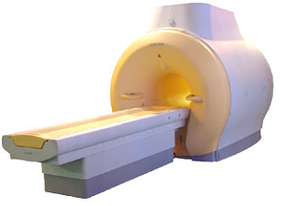
'MRI system is not an expensive equipment anymore.
ENCORE developed by ISOL Technology is a low cost MRI system with the advantages like of the 1.0T MRI scanner. Developed specially for the overseas market, the ENCORE is gaining popularity in the domestic market by medium sized hospitals.
Due to the optimum RF and Gradient application technology. ENCORE enables to obtain high resolution imaging and 2D/3D Angio images which was only possible in high field MR systems.'
- Less consumption of the helium gas due to the ultra-lightweight magnet specially designed and manufactured for ISOL.
- Cost efficiency MR system due to air cooling type (equivalent to permanent magnetic).
- Patient processing speed of less than 20 minutes.'
Device Information and Specification
CLINICAL APPLICATION
Whole body
CONFIGURATION
Short bore compact
| |  | |
• View the DATABASE results for 'ENCORE 0.5T™' (2).
| | | | |
|  |  | Searchterm 'Meter' was also found in the following services: | | | | |
|  |  |
| |
|
Flow phenomena are intrinsic processes in the human body. Organs like the heart, the brain or the kidneys need large amounts of blood and the blood flow varies depending on their degree of activity. Magnetic resonance imaging has a high sensitivity to flow and offers accurate, reproducible, and noninvasive methods for the quantification of flow. MRI flow measurements yield information of blood supply of of various vessels and tissues as well as cerebro spinal fluid movement.
Flow can be measured and visualized with different pulse sequences (e.g. phase contrast sequence, cine sequence, time of flight angiography) or contrast enhanced MRI methods (e.g. perfusion imaging, arterial spin labeling).
The blood volume per time (flow) is measured in: cm3/s or ml/min. The blood flow-velocity decreases gradually dependent on the vessel dia meter, from approximately 50 cm per second in arteries with a dia meter of around 6 mm like the carotids, to 0.3 cm per second in the small arterioles.
Different flow types in human body:
•
Behaves like stationary tissue, the signal intensity depends on T1, T2 and PD = Stagnant flow
•
Flow with consistent velocities across a vessel = Laminar flow
•
Laminar flow passes through a stricture or stenosis (in the center fast flow, near the walls the flow spirals) = Vortex flow
•
Flow at different velocities that fluctuates = Turbulent flow
See also Flow Effects, Flow Artifact, Flow Quantification, Flow Related Enhancement, Flow Encoding, Flow Void, Cerebro Spinal Fluid Pulsation Artifact, Cardiovascular Imaging and Cardiac MRI. | | | |  | |
• View the DATABASE results for 'Flow' (113).
| | |
• View the NEWS results for 'Flow' (7).
| | | | |  Further Reading: Further Reading: | News & More:
|
|
| |
|  |  | Searchterm 'Meter' was also found in the following services: | | | | |
|  |  |
| |
|
A problem occurs in the phase encoding direction, where the phases of signal-bearing tissues outside of the FOV in the y-direction are a replication of the phases that are encoded within the FOV. This signal will be mapped (wrapped, backfolded) back into the image at incorrect locations.
Foldover suppression ( phase oversampling, no phase wrap) is a user-selectable para meter that maps this signal to its correct location outside the FOV, then discards any signal from outside the FOV before displaying the image. In order to be able to choose this para meter, in most cases more than an average is necessary.
See also Phase Wrapping Artifact and Oversampling. | |  | |
• View the DATABASE results for 'Foldover Suppression' (4).
| | | | |
|  | |  |  |
|  | |
|  | | |
|
| |
 | Look
Ups |
| |