Magnetic Resonance - Technology Information Portal Welcome to MRI Technology
Info
  Sheets

Out-
      side


 'FISP' 
SEARCH FOR    
 
  2 3 5 A B C D E F G H I J K L M N O P Q R S T U V W X Y Z
Result : Searchterm 'FISP' found in 0 term [] and 23 definitions []
previous     6 - 10 (of 23)     next
Result Pages : [1 2 3 4 5]
Searchterm 'FISP' was also found in the following service: 
spacer
Forum  (2)  
 
ENCORE 0.5T™InfoSheet: - Devices -
Intro, 
Types of Magnets, 
Overview, 
etc.MRI Resource Directory:
 - Devices -
 
www.isoltech.co.kr/english/product/05t.htm 'MRI system is not an expensive equipment anymore. ENCORE developed by ISOL Technology is a low cost MRI system with the advantages like of the 1.0T MRI scanner. Developed specially for the overseas market, the ENCORE is gaining popularity in the domestic market by medium sized hospitals.
Due to the optimum RF and Gradient application technology. ENCORE enables to obtain high resolution imaging and 2D/3D Angio images which was only possible in high field MR systems.'
- Less consumption of the helium gas due to the ultra-lightweight magnet specially designed and manufactured for ISOL. - Cost efficiency MR system due to air cooling type (equivalent to permanent magnetic). - Patient processing speed of less than 20 minutes.'
Device Information and Specification
CLINICAL APPLICATION
Whole body
CONFIGURATION
Short bore compact
Head, C-spine, L-spine, TMJ, Knee, Shoulder, General purpose, Phased Array System: 4 digital receiver channels (Up to 12 channels)
SYNCHRONIZATION
ECG/peripheral: Optional/yes, respiratory gating
SINGLE/MULTI SLICE
Image reconstruction time (2562 ) : 0.02 s
FOV
40 cm
BORE DIAMETER
or W x H
58 cm diameter
MAGNET WEIGHT
3200 kg
H*W*D
200 x 168 x 187 cm
COOLING SYSTEM TYPE
Air-cooled coil and amplifier
CRYOGEN USE
0.05 L/hr helium
STRENGTH
15 mT/m
5-GAUSS FRINGE FIELD
2.3 m / 3.1 m
Passive and active
spacer
 
• Related Searches:
    • Echelon™ 1.5T
    • Superconducting Magnet
    • Coil
    • Low Field MRI
    • MRI
MRI Resources 
Blood Flow Imaging - MRI Technician and Technologist Career - Calculation - Most Wanted - DICOM - Crystallography
 
MAGNETOM C™InfoSheet: - Devices -
Intro, 
Types of Magnets, 
Overview, 
etc.MRI Resource Directory:
 - Devices -
 
www.medical.siemens.com 
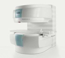
From Siemens Medical Systems;
A new, powerful, compact player in MRI. For both, patients and health care professionals, the mid-field has realized a giant step to cost efficient quality care. Obese patients and people with claustrophobia appreciate the comfortable side loading. The smallest pole diameter - 137 cm (54 inches) allows for optimal patient comfort.
Device Information and Specification
CLINICAL APPLICATION
Whole body
CONFIGURATION
C-shaped open MRI
Multi channel imaging, CP Head//Neck Array Coil, Body/Spine Array Coil, large, Transmit Coil
SYNCHRONIZATION
ECG/peripheral: Optional/yes, respiratory gating
PULSE SEQUENCES
SE, FLASH, FISP, IR, FIR, STIR, TrueIR/FISP, FSE, MT, SS-FSE, MT-SE, MTC, MSE, EPI, PSIF
IMAGING MODES
Single, multislice, volume study, multi angle, multi oblique
TR
Min. TR 2.81 ms
TE
Min. TE 0.98 ms
512 x 512 full screen display
MEASURING MATRIX
64 x 64 to 512 x 512
FOV
0.5 - 40 cm
MAGNET TYPE
Permanent
BORE DIAMETER
or W x H
41 cm vertical gap distance
MAGNET WEIGHT
16000 kg
H*W*D
233 x 206 x 160 cm
STRENGTH
24 mT/m
5-GAUSS FRINGE FIELD
2.2 m / 2.2 m
Passive and active
spacer

• View the DATABASE results for 'MAGNETOM C™' (2).Open this link in a new window

 
Further Reading:
  Basics:
Section 2: 510(k) Summary, MAGNETOM C! System Classification Name: Magnetic Resonance Diagnostic(.pdf)
   by www.accessdata.fda.gov    
MRI Resources 
Online Books - Libraries - Collections - Bioinformatics - MRI Technician and Technologist Jobs - Artifacts
 
Balanced SequenceForum -
related threadsInfoSheet: - Sequences - 
Intro, 
Overview, 
Types of, 
etc.
 
This family of sequences uses a balanced gradient waveform. This waveform will act on any stationary spin on resonance between 2 consecutive RF pulses and return it to the same phase it had before the gradients were applied. A balanced sequence starts out with a RF pulse of 90° or less and the spins in the steady state. Prior to the next TR in the slice encoding, the phase encoding and the frequency encoding direction, gradients are balanced so their net value is zero. Now the spins are prepared to accept the next RF pulse, and their corresponding signal can become part of the new transverse magnetization. If the balanced gradients maintain the longitudinal and transverse magnetization, the result is that both T1 and T2 contrast are represented in the image.
This pulse sequence produces images with increased signal from fluid (like T2 weighted sequences), along with retaining T1 weighted tissue contrast. Balanced sequences are particularly useful in cardiac MRI. Because this form of sequence is extremely dependent on field homogeneity, it is essential to run a shimming prior the acquisition.
Usually the gray and white matter contrast is poor, making this type of sequence unsuited for brain MRI. Modifications like ramping up and down the flip angles can increase signal to noise ratio and contrast of brain tissues (suggested under the name COSMIC - Coherent Oscillatory State acquisition for the Manipulation of Image Contrast).
These sequences include e.g. Balanced Fast Field Echo (bFFE), Balanced Turbo Field Echo (bTFE), Fast Imaging with Steady Precession (TrueFISP, sometimes short TRUFI), Completely Balanced Steady State (CBASS) and Balanced SARGE (BASG).
 
Images, Movies, Sliders:
 Cardiac Infarct Short Axis Cine Overview  Open this link in a new window
    

Courtesy of  Robert R. Edelman
 Infarct 4 Chamber Cine  Open this link in a new window
    

Courtesy of  Robert R. Edelman
 
spacer

• View the DATABASE results for 'Balanced Sequence' (5).Open this link in a new window

 
Further Reading:
  News & More:
Generic Eddy Current Compensation for Rapid Magnetic Resonance Imaging(.pdf)
   by www.switt.ch    
Magnetic resonance imaging guided musculoskeletal interventions at 0.23T: Chapter 4. Materials and methods
2002
Searchterm 'FISP' was also found in the following service: 
spacer
Forum  (2)  
 
CHORUS 1.5T™InfoSheet: - Devices -
Intro, 
Types of Magnets, 
Overview, 
etc.MRI Resource Directory:
 - Devices -
 
www.isoltech.co.kr/english/product/default.htm 'Next generation MRI system 1.5T CHORUS developed by ISOL Technology is optimized for both clinical diagnostic imaging and for research development.
CHORUS offers the complete range of feature oriented advanced imaging techniques- for both clinical routine and research. The compact short bore magnet, the patient friendly design and the gradient technology make the innovation to new degree of perfection in magnetic resonance.'
Device Information and Specification
CLINICAL APPLICATION
Whole body
CONFIGURATION
Short bore
Head, C-spine, L-spine, TMJ, Knee, Shoulder, General purpose, Phased Array System: 4 digital receiver channels (Up to 12 channels)
Optional
SYNCHRONIZATION
ECG/peripheral: Optional/yes, respiratory gating
PULSE SEQUENCES
Spin Echo, Gradient Echo, Fast Spin Echo, Inversion Recovery (STIR, Fluid Attenuated Inversion Recovery), FLASH, FISP, PSIF, Turbo Flash ( MPRAGE ),TOF MR Angiography, Standard echo planar imaging package (SE-EPI, GE-EPI), Optional: Advanced P.A. Imaging Package (up to 4 ch.), Advanced echo planar imaging package, Single Shot and Diffusion Weighted EPI, IR/FLAIR EPI
IMAGING MODES
2D/3D, Travelling Sat, Multi-Slab 3D, MTC and TONE Pulse Sequence, Fat/Water Suppression, Presaturation (up to 6 bands), Flow Compensation using GMR pulse, Multi-Slice, Multi-Group Imaging
SINGLE/MULTI SLICE
Image reconstruction time (2562 ) : 0.02 s
FOV
40 cm
BORE DIAMETER
or W x H
58 cm diameter (patient)
MAGNET WEIGHT
4050 kg
H*W*D
233 x 206 x 160 cm
COOLING SYSTEM TYPE
Water-cooled coil and air-cooled amplifier
CRYOGEN USE
0.07 L/hr helium
STRENGTH
20 mT/m (Upto 27 mT/m)
5-GAUSS FRINGE FIELD
2.5 m / 3.8 m
Passive and active
spacer

• View the DATABASE results for 'CHORUS 1.5T™' (2).Open this link in a new window

MRI Resources 
Pacemaker - DICOM - Journals - Software - Service and Support - Mobile MRI Rental
 
Coronary AngiographyMRI Resource Directory:
 - Cardiovascular Imaging -
 
(MRI-CA, MRCA) The noninvasive imaging of the coronary arteries using magnetic resonance imaging of the heart.
For cardiac MRI-CA, high performance machines are necessary with minimum 40mT/m and 300μsec slew rate.
2D and 3D acquisition are used for fast gradient echo sequences with techniques for minimizing cardiac and respiratory motion and suppressing the high signal of pericardial fat. The optimal sequences seem to be trueFISP, Balanced FFE or FIESTA with SMASH and SENSE techniques. Respiratory motion is minimized for 3D acquisitions by using respiratory gating, especially using navigator echoes (Navigator Technique) to track diaphragmatic and cardiac movement. Optimization of MR technique can provide mapping of long segments of the coronary arteries.
Blood pool agents are being applied to improve the reliability of coronary MR angiography. The major current clinical indication is the identification of coronary artery anomalies because the diagnostic accuracy's for identifying haemodynamically significant stenoses are variable depending of the image quality.

See also Magnetic Resonance Angiography, and Cardiac MRI.
spacer

• View the DATABASE results for 'Coronary Angiography' (7).Open this link in a new window

 
Further Reading:
  Basics:
Role of Magnetic Resonance Imaging in Visualizing Coronary Arteries
Monday, 2 August 2004   by www.clinmedres.org    
  News & More:
Graphic illustration
Tuesday, 12 February 2008   by www.theengineer.co.uk    
MRI Resources 
Fluorescence - MRI Accidents - Knee MRI - Research Labs - Cardiovascular Imaging - MR Myelography
 
previous      6 - 10 (of 23)     next
Result Pages : [1 2 3 4 5]
 Random Page
 
Share This Page
FacebookTwitterLinkedIn

MR-TIP    
Community   
User
Pass
Forgot your UserID/Password ?    


How AI will impact MRI :
only diagnostics 
saving time 
reducing cost 
makes planning obsolete 
reduce human knowledge 
not at all 

Look
      Ups


MR-TIP.com uses cookies! By browsing MR-TIP.com, you agree to our use of cookies.

Magnetic Resonance - Technology Information Portal
Member of SoftWays' Medical Imaging Group - MR-TIP • Radiology-TIP • Medical-Ultrasound-Imaging • 
Copyright © 2003 - 2024 SoftWays. All rights reserved. [ 29 April 2024]
Terms of Use | Privacy Policy | Advertising
 [last update: 2024-02-26 03:41:00]