 | Info
Sheets |
| | | | | | | | | | | | | | | | | | | | | | | | |
 | Out-
side |
| | | | |
|
| | | | | |  |
Result : Searchterm 'fmri' found in 0 term [ ] and 11 definitions [ ] and 11 definitions [ ] ]
| 1 - 5 (of 11) nextResult Pages :  [1 2 3] [1 2 3] |  | |  | Searchterm 'fmri' was also found in the following services: | | | | |
|  |  |
| |
|
(fMRI) Functional magnetic resonance imaging is a technique used to determine the dynamic brain function, often based on echo planar imaging, but can also be performed by using contrast agents and observing their first pass effects through brain tissue. Functional magnetic resonance imaging allows insights in a dysfunctional brain as well as into the basic workings of the brain.
The in functional brain MRI most frequently used effect to assess brain function is the blood oxygenation level dependent contrast ( BOLD) effect, in which differential changes in brain perfusion and their resultant effect on the regional distribution of oxy- to deoxyhaemoglobin are observable because of the different 'intrinsic contrast media' effects of the two haemoglobin forms. Increased brain activity causes an increased demand for oxygen, and the vascular system actually overcompensates for this, increasing the amount of oxygenated haemoglobin. Because deoxygenated haemoglobin attenuates the MR signal, the vascular response leads to a signal increase that is related to the neural activity.
Functional imaging relates body function or thought to specific locations where the neural activity is taking place. The brain is scanned at low resolution but at a fast rate (typically once every 2-3 seconds). Structural MRI together with fMRI provides an anatomical baseline and best spatial resolution.
Interactions can also be seen from the motor cortex to the cerebellum or basal ganglia in the case of a movement disorder such as ataxia. For example: by a finger movement the briefly increase in the blood circulation of the appropriate part of the brain controlling that movement, can be measured. | |  | | | | • Share the entry 'Functional Magnetic Resonance Imaging':    | | | | | | | | | |  Further Reading: Further Reading: | | Basics:
|
|
News & More:
| |
| |
|  | |  |  |  |
| |
|
Special imaging primarily means advanced MRI techniques used for qualitative and quantitative measurement of biological metabolism as e.g., spectroscopy, perfusion imaging (PWI, ASL), diffusion weighted imaging ( DWI, DTI, DTT) and brain function ( BOLD, fMRI). This physiological magnetic resonance techniques offer insights into brain structure, function, and metabolism.
Spectroscopy provides functional information related to identification and quantification of e.g. brain metabolites.
MR perfusion imaging has applications in stroke, trauma, and brain neoplasm. MRI provides the high spatial and temporal resolution needed to measure blood flow to the brain. arterial spin labeling techniques utilize the intrinsic protons of blood and brain tissue, labeled by special preparation pulses, rather than exogenous tracers injected into the blood.
MR diffusion tensor imaging characterizes the ability of water to spread across the brain in different directions. Diffusion parallel to nerve fibers has been shown to be greater than diffusion in the perpendicular direction. This provides a tool to study in vivo fiber connectivity in brain MRI.
FMRI allows the detection of a functional activation in the brain because cortical activity is intimately related to local metabolism changes. See also Diffusion Tensor Tractography. | |  | |
• View the NEWS results for 'Special Imaging' (14).
| | | | |  Further Reading: Further Reading: | Basics:
|
|
News & More:
| |
| |
|  | |  |  |  |
| |
|
Brain imaging, magnetic resonance imaging of the head or skull, cranial magnetic resonance tomography (MRT), neurological MRI - they describe all the same radiological imaging technique for medical diagnostic.
Magnetic resonance imaging of the human brain includes the anatomic description and the detection of lesions. Special techniques like diffusion weighted imaging, functional magnetic resonance imaging ( fMRI) and spectroscopy provide also information about the function and chemical metabolites of the brain.
MRI provides detailed pictures of brain and nerve tissues in multiple planes without obstruction by overlying bones. Brain MRI is the procedure of choice for most brain disorders. It provides clear images of the brainstem and posterior brain, which are difficult to view on a CT scan. It is also useful for the diagnosis of demyelinating disorders (disorders such as multiple sclerosis (MS) that cause destruction of the myelin sheath of the nerve).
With this noninvasive procedure also the evaluation of blood flow and the flow of cerebrospinal fluid (CSF) is possible. Different MRA methods, also without contrast agents can show a venous or arterial angiogram. MRI can distinguish tumors, inflammatory lesions, and other pathologies from the normal brain anatomy. However, MRI scans are also used instead other methods to avoid the dangers of interventional procedures like angiography (DSA - digital subtraction angiography) as well as of repeated exposure to radiation as required for computed tomography (CT) and other X-ray examinations.
A ( birdcage) bird cage coil achieves uniform excitation and reception and is commonly used to study the brain. Usually a brain MRI procedure includes FLAIR, T2 weighted and T1 weighted sequences in two or three planes. See also Fetal MRI, Fluid Attenuation Inversion Recovery ( FLAIR), Perfusion Imaging and High Field MRI. See also Arterial Spin Labeling. | | | | | | | |
• View the DATABASE results for 'Brain MRI' (14).
| | |
• View the NEWS results for 'Brain MRI' (32).
| | | | |  Further Reading: Further Reading: | Basics:
|
|
News & More:
|  |
MRI Reveals Significant Brain Abnormalities Post-COVID
Monday, 21 November 2022 by neurosciencenews.com |  |  |
Combining genetics and brain MRI can aid in predicting chances of Alzheimer's disease
Wednesday, 29 June 2022 by www.sciencedaily.com |  |  |
Roundup: How Even Mild COVID Can Affect the Brain; This Many Daily Steps Improves Longevity; and More
Friday, 11 March 2022 by baptisthealth.net |  |  |
A low-cost and shielding-free ultra-low-field brain MRI scanner
Tuesday, 14 December 2021 by www.nature.com |  |  |
Large International Study Reveals Spectrum of COVID-19 Brain Complications
Tuesday, 9 November 2021 by www.itnonline.com |  |  |
Brain MRI-Based Subtypes of MS Predict Disability Progression, Treatment Response
Thursday, 13 May 2021 by www.neurologyadvisor.com |  |  |
New MRI method improves detection of disease changes in the brain's network
Thursday, 11 June 2020 by www.compute.dtu.dk |  |  |
New NeuroCOVID Classification System Uses MRI to Categorize Patients
Friday, 12 June 2020 by www.diagnosticimaging.com |  |  |
New MRI technique can 'see' molecular changes in the brain
Thursday, 5 September 2019 by medicalxpress.com |  |  |
Talking therapy or medication for depression: Brain scan may help suggest better treatment
Monday, 27 March 2017 by www.newsnation.in |  |  |
MRI identifies brain abnormalities in chronic fatigue syndrome patients
Wednesday, 29 October 2014 by www.eurekalert.org |  |  |
MRIs Useful in Tracking Depression in MS Patients
Tuesday, 1 July 2014 by www.hcplive.com |  |  |
Contrast agent linked with brain abnormalities on MRI
Tuesday, 17 December 2013 by www.sciencecodex.com |  |  |
MRIs Reveal Signs of Brain Injuries Not Seen in CT Scans
Tuesday, 18 December 2012 by www.sciencedaily.com |  |  |
Iron Deposits in the Brain May Be Early Indicator of MS
Wednesday, 13 November 2013 by www.healthline.com |  |  |
Migraine Sufferers Have Thicker Brain Cortex
Tuesday, 20 November 2007 by www.medicalnewstoday.com |
|
| |
|  |  | Searchterm 'fmri' was also found in the following services: | | | | |
|  |  |
| |
|

Swiss-based, formerly Bruker AG - split on the 5th October 2001 into the groups: Bruker Daltonics ( Mass spectroscopy), Bruker Optics (Infrared spectroscopy), Bruker AXS (X-ray spectroscopy) and Bruker BioSpin (The largest part, the NMR business core, the EPR and the Tomography activities).
Product Lines:
•
PharmaScan® - MRI//MRS systems tailored to high-throughput and routine applications in pharmaceutical research.
Product Specification
Contact Information
Please see Bruker BioSpin AG's
| |  | |
• View the NEWS results for 'Bruker BioSpin AG' (1).
| | | | |  Further Reading: Further Reading: | News & More:
|
|
| |
|  | |  |  |  |
| |
|

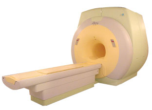
From ISOL Technology
'Ultra high field MR system, it's right close to you.
FORTE 3.0T is the new standard for the future ultra high field MR system.
If you are pushing the limits of your existing clinical MR scanner, the FORTE will surely take you to the next level of diagnostic imaging.
FORTE is the core leader of the medical technology in the 21st century. Proving effects of fMRI that cannot be measured with MRI less than 2.0T.'
Device Information and Specification
CLINICAL APPLICATION
Whole body
CONFIGURATION
Short bore compact
128 x 128, 256 x 256, 512 x 512, 1024 x 1024
| |  | |
• View the DATABASE results for 'FORTE 3.0T™' (2).
| | | | |
|  | |  |  |
|  | 1 - 5 (of 11) nextResult Pages :  [1 2 3] [1 2 3] |
| |
|
| |
 | Look
Ups |
| |