Magnetic Resonance - Technology Information Portal Welcome to MRI Technology
Info
  Sheets

Out-
      side


 'Spin Density' 
SEARCH FOR    
 
  2 3 5 A B C D E F G H I J K L M N O P Q R S T U V W X Y Z
Result : Searchterm 'Spin Density' found in 1 term [] and 8 definitions [], (+ 12 Boolean[] results
previous     11 - 15 (of 21)     next
Result Pages : [1]  [2]  [3 4 5]
MRI Resources 
Non-English - Stimulator pool - Pregnancy - Diffusion Weighted Imaging - Jobs pool - Spectroscopy pool
 
MAGNETOM Skyra
 
www.healthcare.siemens.com/magnetic-resonance-imaging/0-35-to-1-5t-mri-scanner/magnetom-skyra/ 
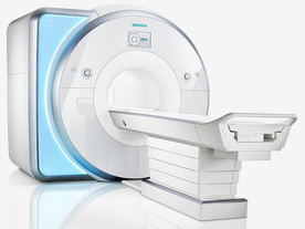
From Siemens Medical Systems; Received FDA clearance in 2010.
MAGNETOM Skyra is a top-of-the-line, patient friendly wide bore 3 Tesla MRI system.
The system is equipped with the Tim 4G and Dot system (Total imaging matrix and Day optimizing throughput), to enhance both productivity and image quality with the complete range of advanced applications for clinical routine and research. Tim 4G features lighter, trimmer MRI coils that take up less space inside the magnet but deliver a high coil element density with increased signal to noise ratio and the possibility to use high iPAT factors.
Device Information and Specification
CLINICAL APPLICATION
Whole Body
CONFIGURATION
Open bore
3 Tesla
Head, spine, torso/ body coil, neurovascular, cardiac, neck, shoulder, knee, wrist, foot//ankle and multi-purpose flex coils. Peripheral vascular, breast, shoulder.
CHANNELS (min. / max. configuration)
48, 64, 128
Chemical shift imaging, single voxel spectroscopy
IMAGING TECHNIQUES
iPAT, mSENSE and GRAPPA (image, k-space),CAIPIRINHA (k-space), noncontrast angiography, plaque imaging, radial motion compensation, Dixon
MINIMUM TR
3D T1 spoiled GRE: 0.95 (256 matrix)
MINIMUM TE
3D T1 spoiled GRE: 0.22 (256 matrix), Ultra-short TE
FOV
0.5 - 50 cm
BORE DIAMETER
or W x H
At isocenter: L-R 70 cm, A-P (with table) 55 cm
TABLE CAPACITY
250 kg
MAGNET WEIGHT (gantry included)
5768 kg
DIMENSION H*W*D (gantry included)
173 x 231 x 219 cm
5-GAUSS FRINGE FIELD
2.6 m / 4.6 m
CRYOGEN USE
Zero boil off rate, approx. 10 years
COOLING SYSTEM
Water; single cryogen, 2 stage refrigeration
up to 200 T/m/s
MAX. AMPLITUDE
45 mT/m
3 linear with 20 coils, 5 nonlinear 2nd-order
POWER REQUIREMENTS
380 / 400 / 420 / 440 / 460 / 480 V, 3-phase + ground; 110 kVA
spacer
MRI Resources 
Non-English - Raman Spectroscopy - Artifacts - General - Safety pool - PACS
 
Lung ImagingMRI Resource Directory:
 - Lung Imaging -
 
Lung imaging is furthermore a challenge in MRI because of the predominance of air within the lungs and associated susceptibility issues as well as low signal to noise of the inflated lung parenchyma. Cardiac and respiratory triggered or breath hold sequences allow diagnostic imaging, however a comparable image quality with computed tomography is still difficult to achieve.
Assumptions for lung MRI:
•
Low signal to noise ratio of the inherently low lung proton density.
•
Cardiac and respiratory motion artifacts.
•
Magnetic susceptibility effects of large magnetic field gradients.
•
Very short transverse relaxation times and significant diffusion yielding short T2 (30-70 msec), short T2* (1-3 msec), and additional long T1 relaxation times (1300-1500 msec).
•
The extreme short T2 values are responsible for a fast signal decay during a single shot readout, resulting in blurring.

The current trends in MRI are the use of new imaging technologies and increasingly powerful magnetic fields. Among these technologies are parallel imaging techniques as well as ventilation agents like hyperpolarized helium for the use as an inert inhalational contrast agent to study lung ventilation properties. With hyperpolarized gases clear images of the lungs can be obtained without using a large magnetic field (see also back projection imaging). Single shot sequences (e.g. TSE or Half Fourier Acquisition Single Shot Turbo Spin Echo HASTE) used in lung MR imaging benefits from parallel imaging techniques due to reduced relaxation time effects during the echo train and therefore reduced image blurring as well as reduced motion artifacts.
In the future, more effective contrast agents may provide an alternative solution to the need for high field MRI. Dynamic contrast enhanced MRI perfusion has demonstrated a potential in the diagnosis of pulmonary embolism or to characterize lung cancer and mediastinal tumors. 3D contrast enhanced magnetic resonance angiography of the thoracic vessel.

See also the related poll result: 'MRI will have replaced 50% of x-ray exams by'
 
Images, Movies, Sliders:
 Anatomic Imaging of the Lungs  Open this link in a new window
      

Courtesy of  Robert R. Edelman
 Normal Lung Gd Perfusion MRI  Open this link in a new window
      

Courtesy of  Robert R. Edelman

 MRI Thorax Basal Plane  Open this link in a new window
 
Radiology-tip.comradLung Scintigraphy
spacer

• View the DATABASE results for 'Lung Imaging' (7).Open this link in a new window


• View the NEWS results for 'Lung Imaging' (3).Open this link in a new window.
 
Further Reading:
  Basics:
A safer approach for diagnostic medical imaging
Monday, 29 September 2014   by www.eurekalert.org    
Parallel Lung Imaging(.pdf)
  News & More:
Chest MRI a viable alternative to chest CT in COVID-19 pneumonia follow-up
Monday, 21 September 2020   by www.healthimaging.com    
CT Imaging Features of 2019 Novel Corona virus (2019-nCoV)
Tuesday, 4 February 2020   by pubs.rsna.org    
Polarean Imaging Phase III Trial Results Point to Potential Improvements in Lung Imaging
Wednesday, 29 January 2020   by www.diagnosticimaging.com    
Low Power MRI Helps Image Lungs, Brings Costs Down
Thursday, 10 October 2019   by www.medgadget.com    
Chest MRI Using Multivane-XD, a Novel T2-Weighted Free Breathing MR Sequence
Thursday, 11 July 2019   by www.sciencedirect.co    
Researchers Review Importance of Non-Invasive Imaging in Diagnosis and Management of PAH
Wednesday, 11 March 2015   by lungdiseasenews.com    
New MRI Approach Reveals Bronchiectasis' Key Features Within the Lung
Thursday, 13 November 2014   by lungdiseasenews.com    
MRI techniques improve pulmonary embolism detection
Monday, 19 March 2012   by medicalxpress.com    
  News & More:
Partnership with VIDA to streamline adoption of advanced MRI of the lungs
Monday, 11 September 2023   by www.itnonline.com    
MRI Resources 
Mobile MRI Rental - Raman Spectroscopy - MRI Technician and Technologist Jobs - Liver Imaging - Coils - Shielding
 
Dual Echo Fast Acquisition Interleaved Spin EchoInfoSheet: - Sequences - 
Intro, 
Overview, 
Types of, 
etc.
 
(DEFAISE) Simultaneously acquired T2 and density weighted echoes in a FSE sequence.

See Fast Spin Echo.
spacer
 
Further Reading:
  Basics:
BASIC PRINCIPLES OF MR IMAGING
   by spinwarp.ucsd.edu    
MRI Resources 
Lung Imaging - Portals - Veterinary MRI - Cochlear Implant - Absorption and Emission - Distributors
 
Double Turbo Spin EchoInfoSheet: - Sequences - 
Intro, 
Overview, 
Types of, 
etc.MRI Resource Directory:
 - Sequences -
 
(DTSE / DE TSE) Simultaneously acquired T2 and density weighted echoes in a TSE sequence.

See also Fast Spin Echo.
spacer
 
Further Reading:
  Basics:
BASIC PRINCIPLES OF MR IMAGING
   by spinwarp.ucsd.edu    
MRI Resources 
Portals - Bioinformatics - Lung Imaging - IR - Guidance - Manufacturers
 
Fast Imaging with Steady State PrecessionInfoSheet: - Sequences - 
Intro, 
Overview, 
Types of, 
etc.
 
(FISP) A fast imaging sequence, which attempts to combine the signals observed separately in the FADE sequence, generally sensitive about magnetic susceptibility artifacts and imperfections in the gradient waveforms. Confusingly now often used to refer to a refocused FLASH type sequence.
This sequence is very similar to FLASH, except that the spoiler pulse is eliminated. As a result, any transverse magnetization still present at the time of the next RF pulse is incorporated into the steady state. FISP uses a RF pulse that alternates in sign. Because there is still some remaining transverse magnetization at the time of the RF pulse, a RF pulse of a degree flips the spins less than a degree from the longitudinal axis. With small flip angles, very little longitudinal magnetization is lost and the image contrast becomes almost independent of T1. Using a very short TE (with TR 20-50 ms, flip angle 30-45°) eliminates T2* effects, so that the images become proton density weighted. As the flip angle is increased, the contrast becomes increasingly dependent on T1 and T2*. It is in the domain of large flip angles and short TR that FISP exhibits vastly different contrast to FLASH type sequences. Used for T1 orthopedic imaging, 3D MPR, cardiography and angiography.
spacer

• View the DATABASE results for 'Fast Imaging with Steady State Precession' (5).Open this link in a new window

 
Further Reading:
  Basics:
MRI techniques improve pulmonary embolism detection
Monday, 19 March 2012   by medicalxpress.com    
MRI Resources 
Examinations - Patient Information - PACS - Journals - Guidance - Jobs pool
 
previous      11 - 15 (of 21)     next
Result Pages : [1]  [2]  [3 4 5]
 Random Page
 
Share This Page
FacebookTwitterLinkedIn

MR-TIP    
Community   
User
Pass
Forgot your UserID/Password ?    


Personalized protocols (age, gender, body habitus, etc.) lead to :
more automated planning 
improved patient comfort 
better diagnostics 
optimized image quality 
nothing 

Look
      Ups


MR-TIP.com uses cookies! By browsing MR-TIP.com, you agree to our use of cookies.

Magnetic Resonance - Technology Information Portal
Member of SoftWays' Medical Imaging Group - MR-TIP • Radiology-TIP • Medical-Ultrasound-Imaging • 
Copyright © 2003 - 2024 SoftWays. All rights reserved. [ 2 May 2024]
Terms of Use | Privacy Policy | Advertising
 [last update: 2024-02-26 03:41:00]