| Info
Sheets |
| | | | | | | | | | | | | | | | | | | | | | | | |
 | Out-
side |
| | | | |
|
| | | | | |  | Searchterm 'Display' was also found in the following services: | | | | |
|  |  |
| |
|
Device Information and Specification CLINICAL APPLICATION Whole body SE, IR, 2D/3D TurboSE, Turbo IR, Dark-Fluid IR, True IR, 2D/3D MEDIC, 2D/3D GRE FLASH, 2D/3D GRE FISP, 2D/3D PSIF, 2D TurboFLASH, 3D MP-RAGE, 3D TurboFLASH, 2D/3D TOF angiography, MTC, TONE with 3D TOF MRA, GMR, LOTA IMAGING MODES Single, multislice, volume study, multi angle, multi oblique178 images/sec at 256 x 256 at 100% FOV1024 x 1024 full screen display POWER REQUIREMENTS 380/400/420/440/480 V Passive, act.; 1st order std./2nd opt. | | | | | |
| | | | | |
| |
|
From Siemens Medical Systems;
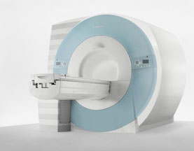
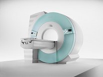
70 cm + 125 cm + 1.5T and Tim - a combination never seen before in MRI ...
MAGNETOM Espree™s unique open bore design can accommodate more types of patients than other 1.5T systems on the market today, in particular the growing population of obese patients. The power of 1.5T combined with Tim technology boosts signal to noise, which is necessary to adequately image obese patients.
Device Information and Specification
CLINICAL APPLICATION
Whole body
Body, Tim [32 x 8], Tim [76 coil elements with up to 18 RF channels])
GRE, IR, FIR, STIR, TrueIR/FISP, FSE, FLAIR, MT, SS-FSE, MT-SE, MTC, MSE, EPI, 3D DESS//CISS/PSIF, GMR
IMAGING MODES
Single, multislice, volume study, multi angle, multi oblique
Image Processor reconstructing up to 3226 images per second (256 x 256, 25% recFoV)
1024 x 1024 full screen display
| | | |
• View the DATABASE results for 'MAGNETOM Espree™' (2).
| | | | |  Further Reading: Further Reading: | News & More:
|
|
| |
| | | | | |
| |
|
( MRA) Magnetic resonance angiography is a medical imaging technique to visualize blood filled structures, including arteries, veins and the heart chambers. This MRI technique creates soft tissue contrast between blood vessels and surrounding tissues primarily created by flow, rather than displaying the vessel lumen. There are bright blood and black blood MRA techniques, named according to the appearance of the blood vessels. With this different MRA techniques both, the blood flow and the condition of the blood vessel walls can be seen. Flow effects in MRI can produce a range of artifacts. MRA takes advantage of these artifacts to create predictable image contrast due to the nature of flow.
Technical parameters of the MRA sequence greatly affect the sensitivity of the images to flow with different velocities or directions, turbulent flow and vessel size.
This are the three main types of MRA:
All angiographic techniques differentially enhance vascular MR signal. The names of the bright blood techniques TOF and PCA reflect the physical properties of flowing blood that were exploited to make the vessels appear bright. Contrast enhanced magnetic resonance angiography creates the angiographic effect by using an intravenously administered MR contrast agent to selectively shorten the T1 of blood and thereby cause the vessels to appear bright on T1 weighted images.
MRA images optimally display areas of constant blood flow-velocity, but there are many situations where the flow within a voxel has non-uniform speed or direction. In a diseased vessel these patterns are even more complex. Similar loss of streamline flow occurs at all vessel junctions and stenoses, and in regions of mural thrombosis. It results in a loss of signal, due to the loss of phase coherence between spins in the voxel.
This signal loss, usually only noticeable distal to a stenosis, used to be an obvious characteristic of MRA images. It is minimized by using small voxels and the shortest possible TE. Signal loss from disorganized flow is most noticeable in TOF imaging but also affects the PCA images.
Indications to perform a magnetic resonance angiography ( MRA):
•
Detection of aneurysms and dissections
•
Evaluation of the vessel anatomy, including variants
•
Blockage by a blood clot or stenosis of the blood vessel caused by plaques (the buildup of fat and calcium deposits)
Conventional angiography or computerized tomography angiography (CT angiography) may be needed after MRA if a problem (such as an aneurysm) is present or if surgery is being considered.
See also Magnetic Resonance Imaging MRI. | | | | | | | | | | |
• View the DATABASE results for 'Magnetic Resonance Angiography MRA' (3).
| | |
• View the NEWS results for 'Magnetic Resonance Angiography MRA' (10).
| | | | | | Further Reading: | | Basics:
|
|
News & More:
| |
| |
| | | Searchterm 'Display' was also found in the following services: | | | | |
| | |
| |
|
The normal image display, at which the grey scale of a display element corresponds to the magnitude of the MR signal. | | | | | |
| | | | | |
| |
|
(PACS) A system used to communicate and archive medical imaging data, mostly images and associated textural data generated in a radiology department, and disseminated throughout the hospital. A PACS is usually based on the DICOM ( Digital Imaging and Communications in Medicine) standard.
The main components in the PACS are:
•
acquisition devices where the images are acquired,
•
short and longer term archives for storage of digital and textural data,
•
a database and database management,
•
diagnostic and review workstations,
•
software to run the system,
•
a communication network linking the system components,
•
interfaces with other networks (hospital and radiological information systems).
Acquisition devices, which acquire their data in direct digital format, like a MRI system, are most easily integrated into a PACS.
Short term archives need to have rapid access, such as provided by a RAID (redundant array of independent disks), whereas long term archives need not have such rapid access and can be consigned, e.g. to optical disks or a magnetic.
High speed networks are necessary for rapid transmission of imaging data from the short term archive to the diagnostic workstations. Optical fiber, ATM (asynchronous transfer mode), fast or switched Ethernet, are examples of high speed transmission networks, whereas demographic textural data may be transmitted along conventional Ethernet.
Sophisticated software is a major element in any hospital-wide PACS. The software concepts include: preloading or prefetching of historical images pertinent to current examinations, worklists and folders to subdivide the vast mass of data acquired in a PACS in a form, which is easy and practical to access, default display protocols whereby images are automatically displayed on workstation monitors in a prearranged clinically logical order and format, and protocols radiologists can rapidly report worklists of undictated examinations, using a minimum of computer manipulation. | | | |
• View the DATABASE results for 'Picture Archiving and Communication System' (5).
| | |
• View the NEWS results for 'Picture Archiving and Communication System' (1).
| | | | | | Further Reading: | Basics:
|
|
| |
| | | | |
| | |
| | | |
|
| |
 | Look
Ups |
| |