Magnetic Resonance - Technology Information Portal Welcome to MRI Technology
Info
  Sheets

Out-
      side


 'Arc' 
SEARCH FOR    
 
  2 3 5 A B C D E F G H I J K L M N O P Q R S T U V W X Y Z
Result : Searchterm 'Arc' found in 6 terms [] and 60 definitions []
previous     21 - 25 (of 66)     next
Result Pages : [1 2]  [3 4 5 6 7 8 9 10 11 12 13 14]
Searchterm 'Arc' was also found in the following services: 
spacer
News  (761)  Resources  (154)  Forum  (48)  
 
MAGNETOM 7T
 
www.healthcare.siemens.com/magnetic-resonance-imaging/7t-mri-scanner/magnetom-7t 
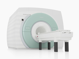
From Siemens Medical Systems;
The MAGNETOM 7T is designed as an open research platform. 7T MRI provides anatomical detail at the submillimiter scale, enhanced contrast mechanisms, outstanding spectroscopy performance, ultra-high resolution functional imaging (fMRI), multinuclear whole-body MRI and functional information.
This ultra high field (UHF) MRI device is a research system and not cleared, approved or licensed in any jurisdiction for patient examinations.
Device Information and Specification
CLINICAL APPLICATION
Whole body
CONFIGURATION
Compact
7 Tesla
High-performance, ultra high field coils available. Integration and support for coil developments.
CHANNELS (min. / max. configuration)
32, optional 8 channels TX array
Chemical shift imaging, single voxel spectroscopy, multinuclear imaging optional
IMAGING TECHNIQUES
iPAT, mSENSE and GRAPPA (image, k-space), noncontrast angiography, plaque imaging, radial motion compensation
FOV
40 x 40 x 30 cm³ less than 8% nonlinearity
BORE DIAMETER
or W x H
60 cm
TABLE CAPACITY
200 kg
MAGNET WEIGHT (gantry included)
35017 kg
DIMENSION H*W*D (gantry included)
320 x 240 x 317,5 cm
5-GAUSS FRINGE FIELD
7.9 m / 5.6 m
CRYOGEN USE
Zero boil off rate
COOLING SYSTEM
Water
up to 200 T/m/s
MAX. AMPLITUDE
up to 70 mT/m
Up to 3rd order shim coils, user configurable B0 shim ? B0 maps and ROI definition
POWER REQUIREMENTS
2000 Volts, 650A
spacer
 
Further Reading:
  Basics:
MAGNETOM 7T Product Brochure
   by www.healthcare.siemens.com    
  News & More:
Ultra-high-field MRI may allow earlier diagnosis of Parkinson's disease
Wednesday, 5 March 2014   by www.sciencedaily.com    
Feasibility of Using Ultra-High Field (7 T) MRI for Clinical Surgical Targeting
Thursday, 17 May 2012   by www.plosone.org    
Ultrahigh-Field MRI May Detect Additional Pathology in EAE
Sunday, 20 October 2013   by www.msdiscovery.org    
Searchterm 'Arc' was also found in the following services: 
spacer
Radiology  (25) Open this link in a new windowUltrasound  (65) Open this link in a new window
MRI EquipmentInfoSheet: - Devices -
Intro, 
Types of Magnets, 
Overview, 
etc.
 
The MRI equipment consists of following components:
The magnet generates the magnetic field.
Shim coils make the magnetic field homogeneous.
Radio frequency coils transmit the radio signal into the body part being imaged.
Receiver coils detect the returning radio signals.
Gradient coils provide spatial localization of the signals.
Shielding coils produce a magnetic field that cancels the field from primary coils in regions where it is not desired.
The computer reconstructs the signals into the image.
The MRI scanner room is shielded by a faraday shield.
Different cooling systems cool the magnet, the scanner room and the technique room.

Better MRI equipment and software design along with the latest information technology improves system maintenance and overall communication. Software and digital imaging and communications in medicine (DICOM) compatibility allows to network into hospital databases, helps to modify pulse sequences, data post processing, and archiving via picture archiving and communication system (PACS).

See also the related poll result: 'Most outages of your scanning system are caused by failure of'
Radiology-tip.comradCT Scanner,  Radiography
spacer
Medical-Ultrasound-Imaging.comUltrasound Machine,  Ultrasound System Performance
spacer

• View the DATABASE results for 'MRI Equipment' (13).Open this link in a new window


• View the NEWS results for 'MRI Equipment' (4).Open this link in a new window.
 
Further Reading:
  News & More:
Low Power MRI Helps Image Lungs, Brings Costs Down
Thursday, 10 October 2019   by www.medgadget.com    
MRI safety targeted as new group offers credentialing test
Monday, 12 January 2015   by www.modernhealthcare.com    
Audio/Video System helps patients relax during MRI scans
Monday, 8 December 2014   by news.thomasnet.com    
Dräger introduces anaesthesia system for MRI environment
Wednesday, 12 December 2007   by www.mtbeurope.info    
MRI Resources 
NMR - Diffusion Weighted Imaging - Journals - Spectroscopy pool - Examinations - MRI Reimbursement
 
Polar Coordinate System
 
A coordinate system, which uses the distance from the coordinate system center and positional angles to identify points in space rather than orthogonal independent unit vectors as in the Cartesian coordinate system. Polar and partly polar (cylindrical) coordinate systems are widely used to describe spin motion in NMR experiments.
It is important to know how to compute the coordinates of a point in the polar coordinate system when they are given in a Cartesian system and vice versa. The length of the vector r pointing from the coordinate origin to a point in 2D space is given as
r = √(x2 + y2).
while the polar or phase angle f is obtained by performing the operation
f = arctan (y/x),
where the arctan function is the inverse of the tangent function.
spacer
Searchterm 'Arc' was also found in the following services: 
spacer
News  (761)  Resources  (154)  Forum  (48)  
 
Radiology Information SystemMRI Resource Directory:
 - RIS -
 
(RIS) Radiology information system means a computer system that stores and processes the information for a radiology department and can be linked to the hospital information system.
The principal purpose of a RIS consists of taking over the general functions of the administration inclusive planning, monitoring and communication of all data regarding patients and its investigations in the radiology. The correct images should reach, at the correct time, the correct users. For this reason the RIS must contain a workflow management in order to simplify and steer the data flow at the individual view stations or devices (laser cameras etc.). The Radiology Information System is optimally complemented with a Picture Archiving and Communication System (PACS).
Collection, storage and administration of patient master data
Archives administration
  • Treatment of requirements
  • Work scheduling
    Account
    Communication (with the hospital information system, MRI scanner, other devices etc.)
    Statistic evaluations
    spacer

    • View the DATABASE results for 'Radiology Information System' (3).Open this link in a new window

     
    Further Reading:
      Basics:
    Healthcare IT Yellow Pages Radiology Information Systems (RIS) Directory
       by www.health-infosys-dir.com    
    Searchterm 'Arc' was also found in the following services: 
    spacer
    Radiology  (25) Open this link in a new windowUltrasound  (65) Open this link in a new window
    Signa 3.0Tâ„¢InfoSheet: - Devices -
Intro, 
Types of Magnets, 
Overview, 
etc.MRI Resource Directory:
 - Devices -
     
    www.gehealthcare.com/usen/mr/s_excite3/index.html (Signa VH/i 3.0T)
    With GE Healthcare leading-edge technology in ultra-high-field imaging. The 3 T VH/i provides a platform for advanced applications in radiology, cardiology, psychology and psychiatry. Real-time image processing lets you acquire multislice whole brain images and map brain functions for research or surgical planning. And the 3 T Signa VH/i is flexible enough to provide clinicians with high performance they require. It can provide not only outstanding features in brain scanning and neuro-system research, but also a wide range of use in scanning breasts, extremities, the spine and the cardiovascular systems.
    Device Information and Specification
    CLINICAL APPLICATION
    Whole body
    CONFIGURATION
    Cylindrical - high homogeneity
    T/R quadrature head, T/R quadrature body, T/R phased array extremity (opt)
    SYNCHRONIZATION
    ECG/peripheral, respiratory gating
    PULSE SEQUENCES
    SE, IR, 2D/3D GRE, FGRE, RF-spoiled GRE, FSE, Angiography: 2D/3D TOF, 2D/3D phase contrast vascular
    IMAGING MODES
    Single, multislice, volume study, fast scan, multi slab, cine, localizer
    SINGLE SLICE
    100 Images/sec with Reflex100
    MULTISLICE
    100 Images/sec with Reflex100
    1 cm to 40 cm continuous
    2D 0.5-100mm in 0.1mm incremental
    1280 x 1024
    MEASURING MATRIX
    128x512 steps 32 phase encode
    PIXEL INTENSITY
    256 gray levels
    55cm
    MAGNET WEIGHT
    15102 kg incl. cryogen's
    H*W*D
    260cm x 238cm x 265cm
    POWER REQUIREMENTS
    480 or 380/415, 3 phase ||
    COOLING SYSTEM TYPE
    Closed-loop water-cooled grad.
    Less than 0.14 L/hr liquid He
    STRENGTH
    40mT/m
    5-GAUSS FRINGE FIELD, radial/axial
    5.4 m x 3.2 m
    Superconductive + hi order active
    spacer

    • View the DATABASE results for 'Signa 3.0T™' (2).Open this link in a new window

    MRI Resources 
    Most Wanted - Implant and Prosthesis pool - - Diffusion Weighted Imaging - Veterinary MRI - Fluorescence
     
    previous      21 - 25 (of 66)     next
    Result Pages : [1 2]  [3 4 5 6 7 8 9 10 11 12 13 14]
     Random Page
     
    Share This Page
    FacebookTwitterLinkedIn

    MR-TIP    
    Community   
    User
    Pass
    Forgot your UserID/Password ?    


    MRI is trending to low field magnets :
    reduced costs will lead to this change 
    AI will close the gap to high field 
    only in remote areas 
    is only temporary 
    never 

    Look
          Ups


    MR-TIP.com uses cookies! By browsing MR-TIP.com, you agree to our use of cookies.

    Magnetic Resonance - Technology Information Portal
    Member of SoftWays' Medical Imaging Group - MR-TIP • Radiology-TIP • Medical-Ultrasound-Imaging • 
    Copyright © 2003 - 2024 SoftWays. All rights reserved. [ 5 May 2024]
    Terms of Use | Privacy Policy | Advertising
     [last update: 2024-02-26 03:41:00]