 | Info
Sheets |
| | | | | | | | | | | | | | | | | | | | | | | | |
 | Out-
side |
| | | | |
|
| | | 'Gradient Echo Multi Slice' | |
Result : Searchterm 'Gradient Echo Multi Slice' found in 1 term [ ] and 0 definition [ ] and 0 definition [ ], (+ 17 Boolean[ ], (+ 17 Boolean[ ] results ] results
| previous 6 - 10 (of 18) nextResult Pages :  [1] [1]  [2 3 4] [2 3 4] |  | | |  |  |  |
| |
|

From GE Healthcare;
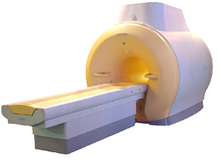
'EXCITE technology has the potential to open the door to new imaging techniques and clinical applications, leaping beyond conventional two and three-dimensional MRI to true 4D imaging that will improve the diagnosis of disease throughout the human body from head to foot.' Robert R. Edelman, M.D., Professor of Radiology at Northwestern University Medical School and Chairman, Department of Radiology, at Evanston Northwestern Healthcare.
Device Information and Specification CLINICAL APPLICATION Whole body Head and body coil standard; all other coils optional; open architecture makes system compatible with a wide selection of coils Optional 2D/3D brain and prostate Standard: SE, IR, 2D/3D GRE and SPGR, Angiography: 2D/3D TOF, 2D/3D Phase Contrast;; 2D/3D FSE, 2D/3D FGRE and FSPGR, SSFP, FLAIR, EPI, optional: 2D/3D Fiesta, FGRET, Spiral, TensorTR 1.3 to 12000 msec in increments of 1 msec TE 0.4 to 2000 msec in increments of 1 msec 2D 0.7 mm to 20 mm; 3D 0.1 mm to 5 mm 128x512 steps 32 phase encode 0.08 mm; 0.02 mm optional POWER REQUIREMENTS 480 or 380/415 less than 0.03 L/hr liquid heliumSTRENGTH SmartSpeed 23 mT/m, HiSpeed Plus 33 mT/m, EchoSpeed Plus 33 mT/m 4.0 m x 2.8 m axial x radial | |  | | | |
|  | |  |  |  |
| |
|

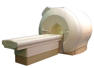
'Next generation MRI system 1.5T CHORUS developed by ISOL Technology is optimized for both clinical diagnostic imaging and for research development.
CHORUS offers the complete range of feature oriented advanced imaging techniques- for both clinical routine and research. The compact short bore magnet, the patient friendly design and the gradient technology make the innovation to new degree of perfection in magnetic resonance.'
Device Information and Specification
CLINICAL APPLICATION
Whole body
Spin Echo, Gradient Echo, Fast Spin Echo,
Inversion Recovery ( STIR, Fluid Attenuated Inversion Recovery), FLASH, FISP, PSIF, Turbo Flash ( MPRAGE ),TOF MR Angiography, Standard echo planar imaging package (SE-EPI, GE-EPI), Optional:
Advanced P.A. Imaging Package (up to 4 ch.), Advanced echo planar imaging package,
Single Shot and Diffusion Weighted EPI, IR/FLAIR EPI
STRENGTH
20 mT/m (Upto 27 mT/m)
| |  | |
• View the DATABASE results for 'CHORUS 1.5T™' (2).
| | | | |
|  | |  |  |  |
| |
|

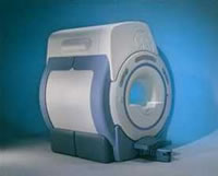
'MRI system is not an expensive equipment anymore.
ENCORE developed by ISOL Technology is a low cost MRI system with the advantages like of the 1.0T MRI scanner. Developed specially for the overseas market, the ENCORE is gaining popularity in the domestic market by medium sized hospitals.
Due to the optimum RF and Gradient application technology. ENCORE enables to obtain high resolution imaging and 2D/3D Angio images which was only possible in high field MR systems.'
- Less consumption of the helium gas due to the ultra-lightweight magnet specially designed and manufactured for ISOL.
- Cost efficiency MR system due to air cooling type (equivalent to permanent magnetic).
- Patient processing speed of less than 20 minutes.'
Device Information and Specification
CLINICAL APPLICATION
Whole body
CONFIGURATION
Short bore compact
| |  | |
• View the DATABASE results for 'ENCORE 0.5T™' (2).
| | | | |
|  | |  |  |  |
| |
|
(2D TOF MRA) This form of MR angiography is based on the acquisition of multiple, short-TR, gradient echo single slice images. 2D TOF MRA is the preferred technique for visualizing slow flow, how for example it happens in veins. 2D TOF MRA consists of multiple sequentially-acquired single slices, therefore the saturation effects are minimized. | |  | | | |
|  | |  |  |  |
| |
|
(DWI) Magnetic resonance imaging is sensitive to diffusion, because the diffusion of water molecules along a field gradient reduces the MR signal. In areas of lower diffusion the signal loss is less intense and the display from this areas is brighter. The use of a bipolar gradient pulse and suitable pulse sequences permits the acquisition of diffusion weighted images (images in which areas of rapid proton diffusion can be distinguished from areas with slow diffusion).
Based on echo planar imaging, multislice DWI is today a standard for imaging brain infarction. With enhanced gradients, the whole brain can be scanned within seconds. The degree of diffusion weighting correlates with the strength of the diffusion gradients, characterized by the b-value, which is a function of the gradient related parameters: strength, duration, and the period between diffusion gradients.
Certain illnesses show restrictions of diffusion, for example demyelinization and cytotoxic edema. Areas of cerebral infarction have decreased apparent diffusion, which results in increased signal intensity on diffusion weighted MRI scans. DWI has been demonstrated to be more sensitive for the early detection of stroke than standard pulse sequences and is closely related to temperature mapping.
DWIBS is a new diffusion weighted imaging technique for the whole body that produces PET-like images. The DWIBS sequence has been developed with the aim to detect lymph nodes and to differentiate normal and hyperplastic from metastatic lymph nodes. This may be possible caused by alterations in microcirculation and water diffusivity within cancer metastases in lymph nodes.
See also Diffusion Weighted Sequence, Perfusion Imaging, ADC Map, Apparent Diffusion Coefficient, and Diffusion Tensor Imaging. | |  | |
• View the DATABASE results for 'Diffusion Weighted Imaging' (11).
| | |
• View the NEWS results for 'Diffusion Weighted Imaging' (4).
| | | | |  Further Reading: Further Reading: | | Basics:
|
|
News & More:
| |
| |
|  | |  |  |
|  | |
|  | | |
|
| |
 | Look
Ups |
| |