 | Info
Sheets |
| | | | | | | | | | | | | | | | | | | | | | | | |
 | Out-
side |
| | | | |
|
| | | | |
Result : Searchterm 'Ultra' found in 4 terms [ ] and 80 definitions [ ] and 80 definitions [ ] ]
| | 1 - 5 (of 84) nextResult Pages : [1] [2 3 4 5 6 7 8 9 10 11 12 13 14 15 16 17] |  | | | | |  |
| |
|
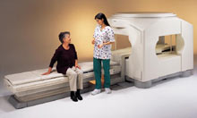
From Toshiba America Medical Systems Inc.;
the Ultra™ system was developed to help healthcare providers be more competitive by delivering greater patient comfort and a broad range of clinical capabilities, says Anita Bowler, product manager, MRI Business Unit, Toshiba America Medical Systems Inc With its unique, powerful gradient technology, the Ultra™ performs advanced clinical studies and consistently provides high-resolution images that are typically associated with high field MRI systems. At the same time, the Ultra™ offers a truly open feeling that makes patients more relaxed, especially those with claustrophobic tendencies.
Device Information and Specification CLINICAL APPLICATION Whole Body Quadrature, solenoid and multi-channel configurations SE, FE, IR, FastSE, FastIR, FastFLAIR, Fast STIR, FastFE, FASE, Hybrid EPI, Multi Shot EPI, Single shot EPI diffusion, True SSFP, SuperFASE; Angiography: 2D(gate/non-gate)/3D TOF, SORS-STC, Black Blood MRA POWER REQUIREMENTS 380/400/415/440/480 V COOLING SYSTEM TYPE Cryogenless | | | | | | • Share the entry 'Ultra™':    | | | | |
 |
| |
|
| | | | | |  Further Reading: Further Reading: | News & More:
|
|
| |
| | | | | |
| |
|
In simple ultrafast GRE imaging, TR and TE are so short, that tissues have a poor imaging signal and - more importantly - poor contrast except when contrast media enhanced ( contrast enhanced angiography). Therefore, the magnetization is 'prepared' during the preparation module, most frequently by an initial 180° inversion pulse.
In the pulse sequence timing diagram, the basic ultrafast gradient echo sequence is illustrated. The 180° inversion pulse is executed one time (to the left of the vertical line), the right side represents the data collection period and is often repeated depending on the acquisition parameters.
See also Pulse Sequence Timing Diagram, there you will find a description of the components.
Ultrafast GRE sequences have a short TR,TE, a low flip angle and TR is so short that image acquisition lasts less than 1 second and typically less than 500 ms. Common TR: 3-5 msec, TE: 2 msec, and the flip angle is about 5°.
Such sequences are often labeled with the prefix 'Turbo' like TurboFLASH, TurboFFE and TurboGRASS.
This allows one to center the subsequent ultrafast GRE data acquisition around the inversion time TI, where one of the tissues of interest has very little signal as its z-magnetization is passing through zero.
Unlike a standard inversion recovery (IR) sequence, all lines or a substantial segment of k-space image lines are acquired after a single inversion pulse, which can then together be considered as readout module. The readout module may use a variable flip angle approach, or the data acquisition may be divided into multiple segments (shots). The latter is useful particularly in cardiac imaging where acquiring all lines in a single segment may take too long relative to the cardiac cycle to provide adequate temporal resolution.
If multiple lines are acquired after a single pulse, the pulse sequence is a type of gradient echo echo planar imaging (EPI) pulse sequence. See also Magnetization Prepared Rapid Gradient Echo ( MPRAGE) and Turbo Field Echo ( TFE). | | | |
• View the DATABASE results for 'Ultrafast Gradient Echo Sequence' (13).
| | | | |
| | | | | |
| |
|
( USPIO) The class of the ultrasmall superparamagnetic iron oxide includes several chemically and pharmacologically very distinct materials, which may or may not be interchangeable for a specific use. Some ultrasmall SPIO particles (median diameter less than 50nm) are used as MRI contrast agents ( Sinerem®, Combidex®), e.g. to differentiate metastatic from inflammatory lymph nodes. USPIO shows also potential for providing important information about angiogenesis in cancer tumors and could possibly complement MRI helping physicians to identify dangerous arteriosclerosis plaques.
Because of the disadvantageous large T2*//T1 ratio, USPIO compounds are less suitable for arterial bolus contrast enhanced magnetic resonance angiography than gadolinium complexes. The tiny ultrasmall superparamagnetic iron oxides do not accumulate in the RES system as fast as larger particles, which results in a long plasma half-life.
USPIO particles, with a small median diameter (less than 10 nm), will accumulate in lymph nodes after an intravenous injection by e.g. direct transcapillary passage through endothelial venules. Once within the nodal parenchyma, phagocytic cells of the mononuclear phagocyte system take up the particles.
As a second way, USPIOs are subsequently taken up from then interstitium by lymphatic vessels and transported to regional lymph nodes. A lymph node with normal phagocytic function takes up a considerable amount and shows a reduction of the signal intensity caused by T2 shortening effects and magnetic susceptibility. Caused by the small uptake of the USPIOs in metastatic lymph nodes, they appear with less signal reduction, and permit the differentiation of healthy lymph nodes from normal-sized, metastatic nodes.
See also Superparamagnetic Contrast Agents, Superparamagnetic Iron Oxide, Very Small Superparamagnetic Iron Oxide Particles, Blood Pool Agents, Intracellular Contrast Agents. | | | |
• View the DATABASE results for 'Ultrasmall Superparamagnetic Iron Oxide' (16).
| | |
• View the NEWS results for 'Ultrasmall Superparamagnetic Iron Oxide' (2).
| | | | | | Further Reading: | | Basics:
|
|
News & More:
| |
| |
| | | | |  |
| |
|
Ultrasound imaging is the primary fetal monitoring modality during pregnancy, nevertheless fetal MRI is increasingly used to image anatomical regions and structures difficult to see with sonography. Given its long record of safety, utility, and cost-effectiveness, ultrasound will remain the modality of first choice in fetal screening. However, MRI is beginning to fill a niche in situations where ultrasound does not provide enough information to diagnose abnormalities before the baby's birth. Magnetic resonance imaging of the fetus provides multiplanar views also in sub-optimal positions, better characterization of anatomic details of e.g. the fetal brain, and information for planning the mode of delivery and airway management at birth.
Indications:
•
Examinations of the placenta
Modern fetal MRI requires no sedatives or muscle relaxants to control fetal movement. Ultrafast MRI techniques (e.g., single shot techniques like Half Fourier Acquisition Single shot Turbo spin Echo HASTE) enable images to be acquired in less than one second to eliminate fetal motion. Such technology has led to increased usage of fetal MRI, which can lead to earlier diagnosis of conditions affecting the baby and has proven useful in planning fetal surgery and designing postnatal treatments. As MR technology continues to improve, more advances in the prenatal diagnosis and treatment of fetal abnormalities are to expect. More advances in in-utero interventions are likely as well. Eventually, fetal MRI may replace even some prenatal tests that require invasive procedures such as amniocentesis.
For Ultrasound Imaging (USI) see Fetal Ultrasound at Medical-Ultrasound-Imaging.com. | | | | | |
• View the DATABASE results for 'Fetal MRI' (5).
| | |
• View the NEWS results for 'Fetal MRI' (2).
| | | | | | Further Reading: | | Basics:
|
|
News & More:
|  |
Advances in medical imaging enable visualization of white matter tracts in fetuses
Wednesday, 12 May 2021 by www.eurekalert.or | | |
Fetal CMR Detects Congenital Heart Defects, Changes Treatment Decisions
Monday, 29 March 2021 by www.diagnosticimaging.com | | |
MRI scans more precisely define and detect some abnormalities in unborn babies
Friday, 12 March 2021 by www.eurekalert.org | | |
Ultrasound and Magnetic Resonance Imaging of Agenesis of the Corpus Callosum in Fetuses: Frontal Horns and Cavum Septi Pellucidi Are Clues to Earlier Diagnosis
Monday, 29 June 2020 by pubmed.ncbi.nlm.nih.gov | | |
MRI helps predict preterm birth
Tuesday, 15 March 2016 by www.eurekalert.org | | |
3-T MRI advancing on ultrasound for imaging fetal abnormalities
Monday, 20 April 2015 by www.eurekalert.org | | |
Babies benefit from pioneering 'miniature' MRI scanner in Sheffield
Friday, 24 January 2014 by www.telegraph.co.uk | | |
Ultrasensitive Detector Pinpoints Big Problem in Tiny Fetal Heart
Tuesday, 6 April 2010 by www.sciencedaily.com | | |
Real-time MRI helps doctors assess beating heart in fetus
Thursday, 29 September 2005 by www.eurekalert.org |
|
| |
| | | | |
| | | |
|
| |
 | Look
Ups |
| |


