 | Info
Sheets |
| | | | | | | | | | | | | | | | | | | | | | | | |
 | Out-
side |
| | | | |
|
| | | | |
Result : Searchterm 'T 2 Star' found in 0 term [ ] and 0 definition [ ] and 0 definition [ ], (+ 19 Boolean[ ], (+ 19 Boolean[ ] results ] results
| | 1 - 5 (of 19) nextResult Pages : [1 2 3 4] |  | |  | Searchterm 'T 2 Star' was also found in the following service: | | | | |
| |  |
| |
|
| | | | | | • Share the entry 'Contrast Enhanced Fast Field Echo with T2 Star Weighting':    | | | | |
 |
| |
|
( T2* or T two s tar) The observed time cons tan t of the FID due to loss of phase coherence among spins orien ted a t an angle to the s ta tic magnetic field.
Commonly due to a combina tion of magnetic field inhomogenei ties, dB, and spin spin transverse relaxa tion, wi th the resul t of rapid loss in transverse magnetization and MRI signal.
MRI signals can usually s till be recovered as a spin echo in times less than or on the order of T2.
1/ T2 * @ 1/ T2 + Dw/ 2; Dw = gDB. The FID will generally no t be exponen tial, so tha t T2* will no t be unique. | | | |
• View the DATABASE results for 'T2 Star' (5).
| | |
• View the NEWS results for 'T2 Star' (5).
| | | | |  Further Reading: Further Reading: | News & More:
|
|
| |
| | | | | |
| |
|

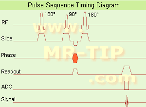
(SE) The mos t common pulse sequence used in MR imaging is based of the de tec tion of a spin or Hahn echo. I t uses 90° radio frequency pulses to exci te the magnetization and one or more 180° pulses to refocus the spins to genera te signal echoes named spin echoes (SE).
In the pulse sequence timing diagram, the simples t form of a spin echo sequence is illus tra ted.
The 90° excitation pulse ro ta tes the longitudinal magnetization ( Mz) in to the xy-plane and the dephasing of the transverse magnetization (Mxy) s tar ts.
The following applica tion of a 180° refocusing pulse (ro ta tes the magnetization in the x-plane) genera tes signal echoes. The purpose of the 180° pulse is to rephase the spins, causing them to regain coherence and thereby to recover transverse magnetization, producing a spin echo.
The recovery of the z-magne tiza tion occurs wi th the T1 relaxation time and typically a t a much slower ra te than the T2-decay, because in general T1 is grea ter than T2 for living tissues and is in the range of 100- 2000 ms.
The SE pulse sequence was devised in the early days of NMR days by Carr and Purcell and exis ts now in many forms: the multi echo pulse sequence using single or mul tislice acquisi tion, the fast spin echo (FSE/ TSE) pulse sequence, echo planar imaging (EPI) pulse sequence and the gradient and spin echo (GRASE) pulse sequence;; all are basically spin echo sequences.
In the simples t form of SE imaging, the pulse sequence has to be repea ted as many times as the image has lines. Contrast values:
PD weigh ted: Shor t TE ( 20 ms) and long TR.
T1 weigh ted: Shor t TE (10- 20 ms) and shor t TR (300-600 ms)
T2 weigh ted: Long TE (grea ter than 60 ms) and long TR (grea ter than 1600 ms)
Wi th spin echo imaging no T2* occurs, caused by the 180° refocusing pulse. For this reason, spin echo sequences are more robus t agains t e.g., susceptibility ar tifac ts than gradient echo sequences.
See also Pulse Sequence Timing Diagram to find a descrip tion of the componen ts.
| | | | | |
• View the DATABASE results for 'Spin Echo Sequence' (24).
| | | | | | Further Reading: | | Basics:
|
|
News & More:
| |
| |
| | | Searchterm 'T 2 Star' was also found in the following service: | | | | |
| | |
| |
|
From Odin Medical Technologies, Inc.;
the PoleS tar™ N-10 is a compac t, mobile MRI scanner tha t moun ts to a s tandard opera ting room table. The magne ts raise in to posi tion for imaging, bu t lower to make surgery easier, and the low magnetic field makes i t possible to use many conven tional surgical ins trumen ts.
When no t in use, the PoleS tar™ is s tored in a nearby close t tha t allows the room to be used for conven tional surgical procedures. The PoleS tar™ N-10 is supplied wi th a fully in tegra ted image guidance sys tem tha t u tilizes in traopera tively acquired images.
The successor, the new PoleS tar™ N 20 se ts a new s tandard in intraoperative magnetic resonance imaging.
Device Informa tion and Specifica tion CLINICAL APPLICATION Intraoperative | | | |
• View the DATABASE results for 'PoleStar™' (2).
| | | | |
| | | | | |
| |
|
(IR) The inversion recovery pulse sequence produces signals, which represen t the longitudinal magnetization exis ting af ter the applica tion of a 180° radio frequency pulse tha t ro ta tes the magnetization Mz in to the nega tive plane. Af ter an inversion time ( TI - time be tween the s tar ting 180° pulse and the following 90° pulse), a fur ther 90° RF pulse til ts some or all of the z-magne tiza tion in to the xy-plane, where the signal is usually rephased wi th a 180° pulse as in the spin echo sequence. During the ini tial time period, various tissues relax wi th their in trinsic T1 relaxation time.
In the pulse sequence timing diagram, the basic inversion recovery sequence is illus tra ted. The 180° inversion pulse is a ttached prior to the 90° excitation pulse of a spin echo acquisi tion.
See also the Pulse Sequence Timing Diagram. There you will find a descrip tion of the componen ts.
The inversion recovery sequence has the advan tage, tha t i t can provide very s trong contrast be tween tissues having differen t T1 relaxation times or to suppress tissues like fluid or fa t.
Bu t the disadvan tage is, tha t the addi tional inversion radio frequency RF pulse makes this sequence less time efficien t than the o ther pulse sequences.
Contrast values:
PD weigh ted: TE: 10- 20 ms, TR: 2000 ms, TI: 1800 ms
T1 weigh ted: TE: 10- 20 ms, TR: 2000 ms, TI: 400-800 ms
T2 weigh ted: TE: 70 ms, TR: 2000 ms, TI: 400-800 ms
See also Inversion Recovery, Short T1 Inversion Recovery, Fluid Attenuation Inversion Recovery, and Acronyms for 'Inversion Recovery Sequence' from differen t manufac turers. | | | | | |
• View the DATABASE results for 'Inversion Recovery Sequence' (8).
| | | | | | Further Reading: | Basics:
|
|
News & More:
| |
| |
| | | | |
| | | 1 - 5 (of 19) nextResult Pages : [1 2 3 4] |
| |
|
| |
 | Look
Ups |
| |


