 | Info
Sheets |
| | | | | | | | | | | | | | | | | | | | | | | | |
 | Out-
side |
| | | | |
|
| | | | |
Result : Searchterm 'R1' found in 1 term [ ] and 39 definitions [ ] and 39 definitions [ ] ]
| | previous 31 - 35 (of 40) nextResult Pages : [1] [2 3 4 5 6 7 8] |  | |  | Searchterm 'R1' was also found in the following services: | | | | |
| |  |
| |
|
The ability of magnetic compounds to increase the relaxation
rates of the surrounding water proton spins.
Relaxivity is used to improve the contrast of the image, and
to study tissue specific areas where the contrast agent
better diffuses or to perform functional magnetic resonance imaging.
The relaxivity of MRI contrast agents depends on the
molecular structure and kinetic of the complex.
To increase the number of water molecules that are in the inner sphere
of the complex, or to slow down the molecular rotational
correlation time, are possibilities to improve the water relaxivity.
Relaxivity units ( r1, r2) are mM -1 * sec -1 (at varying temperatures). | | | | | | | | | | |  Further Reading: Further Reading: | News & More:
|
|
| |
| | | Searchterm 'R1' was also found in the following service: | | | | |
| | |
| |
|
Resovist® is an organ-specific MRI contrast agent, used for the detection and characterization of especially small focal liver lesions.
Resovist® consists of superparamagnetic iron oxide ( SPIO) nanoparticles coated with carboxydextran, which are accumulated by phagocytosis in cells of the reticuloendothelial system (RES) of the liver. The uptake of Resovist® Injection in the reticuloendothelial cells results in a decrease of the signal intensity of normal liver parenchyma on both T2- and T1 weighted images.
Most malignant liver tumors do not contain RES cells and therefore do not uptake the iron particles. The resulting imaging effect is an improved contrast between the tumor (bright) and the surrounding tissue (dark).
Resovist® can be injected as an intravenous bolus, which allows immediate imaging of the liver and reduces the overall examination time. A dynamic imaging strategy after bolus injection supports to characterize lesions.
In comprehensive clinical trials, it demonstrated an excellent safety profile.
In 2001, Resovist® was approved for the European market.
See also Superparamagnetic Iron Oxide.
Resovist® competed with Primovist™, the other liver imaging agent of Bayer Schering Pharma AG. Due to this reason, the production of Resovist® has been abandoned in 2009. Drug Information and Specification T2/T1, Predominantly negative enhancement PHARMACOKINETIC RES-directed CONCENTRATION 0.5 mol Fe/L DOSAGE Less than 60 kg = 0.9 ml, greater than 60 kg = 1.4 ml PREPARATION Finished product PRESENTATION
Pre-filled syringes of 0.9 and 1.4 mL DO NOT RELY ON THE INFORMATION PROVIDED HERE, THEY ARE
NOT A SUBSTITUTE FOR THE ACCOMPANYING PACKAGE INSERT! Distribution Information TERRITORY TRADE NAME DEVELOPMENT
STAGE DISTRIBUTOR Japan Resovist® approved - Australia Resovist® Approved - | | | |
• View the DATABASE results for 'Resovist®' (6).
| | | | | | Further Reading: | News & More:
|
|
| |
| | | | | |
| |
|

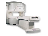
From GE Healthcare;
three dedicated MRI systems - a neuro imager, a cardiovascular system, and a whole body scanner - all in one system.
Device Information and Specification CLINICAL APPLICATION Whole body Head and body coil standard; all other coils optional; open architecture makes system compatible with a wide selection of coils Optional 2D/3D brain and prostate Standard: SE, IR, 2D/3D GRE and SPGR, Angiography: 2D/3D TOF, 2D/3D Phase Contrast;; 2D/3D FSE, 2D/3D FGRE and FSPGR, SSFP, FLAIR, EPI, optional: 2D/3D Fiesta, FGRET, Spiral, TensorTR 1.2 to 12000 msec in increments of 1 msec TE 0.4 to 2000 msec in increments of 1 msec 2D 0.7 mm to 20 mm; 3D 0.1 mm to 5 mm 128x512 steps 32 phase encode POWER REQUIREMENTS 480 or 380/415 Less than 0.03 L/hr liquid He STRENGTH Zoom 40 mT/m, whole body 23 mT/m 4.0 m x 2.8 m axial x radial | | | |
• View the DATABASE results for 'Signa Infinity 1.5T™ TwinSpeed with Excite' (2).
| | | | | | Further Reading: | News & More:
|
|
| |
| | | Searchterm 'R1' was also found in the following services: | | | | |
| | |
| |
|

From GE Healthcare;
'EXCITE technology has the potential to open the door to new imaging techniques and clinical applications, leaping beyond conventional two and three-dimensional MRI to true 4D imaging that will improve the diagnosis of disease throughout the human body from head to foot.' Robert R. Edelman, M.D., Professor of Radiology at Northwestern University Medical School and Chairman, Department of Radiology, at Evanston Northwestern Healthcare.
Device Information and Specification CLINICAL APPLICATION Whole body Head and body coil standard; all other coils optional; open architecture makes system compatible with a wide selection of coils Optional 2D/3D brain and prostate Standard: SE, IR, 2D/3D GRE and SPGR, Angiography: 2D/3D TOF, 2D/3D Phase Contrast;; 2D/3D FSE, 2D/3D FGRE and FSPGR, SSFP, FLAIR, EPI, optional: 2D/3D Fiesta, FGRET, Spiral, TensorTR 1.3 to 12000 msec in increments of 1 msec TE 0.4 to 2000 msec in increments of 1 msec 2D 0.7 mm to 20 mm; 3D 0.1 mm to 5 mm 128x512 steps 32 phase encode 0.08 mm; 0.02 mm optional POWER REQUIREMENTS 480 or 380/415 less than 0.03 L/hr liquid heliumSTRENGTH SmartSpeed 23 mT/m, HiSpeed Plus 33 mT/m, EchoSpeed Plus 33 mT/m 4.0 m x 2.8 m axial x radial | | | |
• View the DATABASE results for 'Signa Infinity 1.5T™ with Excite' (2).
| | | | |
| | | Searchterm 'R1' was also found in the following service: | | | | |
| | |
| |
|
From GE Healthcare;
a friendly and less confining appearance targets the 7% of individuals who refuse to have an MRI because of claustrophobia. This open MRI system is also up to three times faster than other scanners, therefore the Signa OpenSpeed™ reducing exam time and scheduling
issues. In addition, a swing table provides better access and supports up to 500 pounds.
Device Information and Specification CLINICAL APPLICATION Whole body Standard: SE, IR, 2D/3D GRE and SPGR, Angiography: 2D/3D TOF, 2D/3D Phase Contrast;; 2D/3D FSE, 2D/3D FGRE and FSPGR, SSFP, FLAIR, EPI, optional: 2D/3D Fiesta, FGRET, Spiral, TensorTR 1.3 to 12000 msec in increments of 1 msec TE 0.4 to 2000 msec in increments of 1 msec 2D: 0.8mm - 20mm 3D: 0.1mm - 20mm 0.08 mm; 0.02 mm optional POWER REQUIREMENTS 200 - 480, 3-phase | | | |
• View the DATABASE results for 'Signa OpenSpeed™' (2).
| | | | | | Further Reading: | News & More:
|
|
| |
| | | | |
| | |
| | | |
|
| |
 | Look
Ups |
| |


