Diffusion weighted imaging can be performed similar to the phase contrast angiographysequence. The gradients must be increased in amplitude to depict the much slower motions of molecular diffusion in the body.
While a T1 weightedMRIpulse sequence is diffusion sensitive, a quantitative diffusionpulse sequence was introduced by Steijskal and Tanner. Its characteristic features are two strong symmetrical gradient lobes placed on either side of the 180° refocusing pulse in a spin echosequence. These symmetrical gradient lobes have the sole purpose of enhancing dephasing of spins, thereby accelerating intravoxel incoherent motion (IVIM) signal loss.
Dephasing is proportional to the square of the time (diffusion time) during which the gradients are switched on and the strength of the applied gradient field. Therefore, the use of high field gradient systems with faster and more sensitive sequences, make diffusion weighting more feasible.
Areas in which the protons diffuse rapidly (swollen cells in early stroke, less restriction to diffusion) will show an increased signal when the echo is measured relative to areas in which diffusion is restricted.
For increased accuracy of diffusion measurement and image enhancement, useful motion correction techniques such as navigator echo and other methods should be used. In addition to this, applying the b-value calculated by the strength and duration of motion probing gradients with a high rate of accuracy is very important.
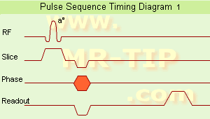
The second picture shows a timing diagram for a 3D pulse sequence. Volume excitation and signal detection are repeated in duration, relative timing and amplitude, each time the sequence is repeated. Two phase encoding components are present, one in the phase encoding direction and the other in slice selection direction (irrespectively incremented in amplitude) in each time the sequence is executed.
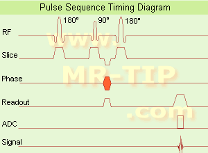
A description of the comparison of hardware activity between different pulse sequences.


 (IR) The

 The schematic figures of a
 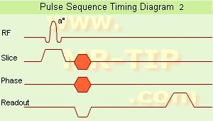 The
The 