Magnetic Resonance - Technology Information Portal Welcome to MRI Technology
Info
  Sheets

Out-
      side


 'Respiratory Trigger' 
SEARCH FOR    
 
  2 3 5 A B C D E F G H I J K L M N O P Q R S T U V W X Y Z
Result : Searchterm 'Respiratory Trigger' found in 1 term [] and 3 definitions [], (+ 4 Boolean[] results
previous     6 - 8 (of 8)     
Result Pages : [1]  [2]
MRI Resources 
Software - Hospitals - Pathology - Mobile MRI - Abdominal Imaging - Research Labs
 
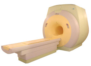
FORTE 3.0Tâ„¢InfoSheet: - Devices -
Intro, 
Types of Magnets, 
Overview, 
etc.MRI Resource Directory:
 - Devices -
 
www.isoltech.co.kr/english/product/30t.htm From ISOL Technology
'Ultra high field MR system, it's right close to you. FORTE 3.0T is the new standard for the future ultra high field MR system. If you are pushing the limits of your existing clinical MR scanner, the FORTE will surely take you to the next level of diagnostic imaging. FORTE is the core leader of the medical technology in the 21st century. Proving effects of fMRI that cannot be measured with MRI less than 2.0T.'
Device Information and Specification
CLINICAL APPLICATION
Whole body
CONFIGURATION
Short bore compact
SYNCHRONIZATION
ECG/peripheral: Optional/yes, external trigger, respiratory gating
PULSE SEQUENCES
Spin echo, Gradient echo, Fast spin echo, Inversion recovery, 2D/3D Fast gradient echo sequences FLAIR/STIR, 2D/3D TOF
IMAGING MODES
2D/3D, T1, T2 and Diffusion//Perfusion imaging, MR Angiography package, Advanced EPI package, Multi-nuclei MR Spectroscopy package
FOV
40 cm
128 x 128, 256 x 256, 512 x 512, 1024 x 1024
BORE DIAMETER
or W x H
61 cm without body coil
MAGNET WEIGHT
12000 kg
H*W*D
260 x 220 x 235 cm
COOLING SYSTEM TYPE
Water-cooled coil and air-cooled amplifier
CRYOGEN USE
0.15 L/hr helium
STRENGTH
38 mT/m
5-GAUSS FRINGE FIELD
3.3 m / 5.2 m
Passive and active
spacer
 
• Related Searches:
    • High Field MRI
    • Device
    • Magnet
    • MRI
    • MRI Equipment
MRI Resources 
Blood Flow Imaging - Pathology - Artifacts - Services and Supplies - Absorption and Emission - Mobile MRI
 
Ghosting ArtifactInfoSheet: - Artifacts - 
Case Studies, 
Reduction Index, 
etc.MRI Resource Directory:
 - Artifacts -
 
Quick Overview
Artifact Information
NAME
Ghosting, ghost
DESCRIPTION
Displaced reduplications of image in phase encoding direction
REASON
Motion, heartbeat, respiration
HELP
Triggering, breath hold, pharmaceuticals to reduce bowel motion
Ghosting artifacts are in the most cases caused by movements (e.g., respiratory motion, bowel motion, arterial pulsations, swallowing, and heartbeat) and appear in the phase encoding direction.
mri safety guidance
Image Guidance
Ghosting artifacts can be reduced by respiratory and cardiac triggering, the use of breath holding pulse sequences, flow compensation or presaturation pulses, depending on their origin. To reduce bowel motion also pharmaceuticals, such as glucagon or scopolamine are useful. This will decrease artifacts from both peristalsis and breathing.

See also Motion Artifact, Phase Encoded Motion Artifact, Cardiac Motion Artifact, and Artifact Reduction - Motion.
spacer

• View the DATABASE results for 'Ghosting Artifact' (5).Open this link in a new window

 
Further Reading:
  Basics:
MRI Artifact Gallery
   by chickscope.beckman.uiuc.edu    
MRI Resources 
MRI Training Courses - Bioinformatics - Liver Imaging - Pediatric and Fetal MRI - Directories - Anatomy
 
Motion ArtifactInfoSheet: - Artifacts - 
Case Studies, 
Reduction Index, 
etc.MRI Resource Directory:
 - Artifacts -
 
Quick Overview
Please note that there are different common names for this artifact.
Artifact Information
NAME
Motion, phase encoded motion, instability, smearing
DESCRIPTION
Blurring and ghosting
REASON
Movement of the imaged object
HELP
Compensation techniques, more averages, anti spasmodic
Patient motion is the largest physiological effect that causes artifacts, often resulting from involuntary movements (e.g. respiration, cardiac motion and blood flow, eye movements and swallowing) and minor subject movements.
Movement of the object being imaged during the sequence results in inconsistencies in phase and amplitude, which lead to blurring and ghosting. The nature of the artifact depends on the timing of the motion with respect to the acquisition. Causes of motion artifacts can also be mechanical vibrations, cryogen boiling, large iron objects moving in the fringe field (e.g. an elevator), loose connections anywhere, pulse timing variations, as well as sample motion. These artifacts appear in the phase encoding direction, independent of the direction of the motion.
mri safety guidance
Image Guidance
Motion artifacts can be flipped 90° by swapping the phase//frequency encoding directions.
The artifacts can be reduced by using breath holding, cardiac synchronization or respiratory compensation techniques: triggering, gating, retrospective triggering or phase encoding artifact reduction. Flow effects can be reduced by using gradient moment nulling of the first order of flow, gradient moment rephasing or flow compensation, depending of the MRI system.
Peristaltic motion can be reduced with the intravenous injection of an anti-spasmodic (e.g. Buscopan).
By using multiple averages, respiratory motion can be reduced in the same way that multiple averages increase the signal to noise ratio. Noticeable motion averaging is seen when four averages are obtained, six averages are often as good as respiratory compensation techniques and higher averages will continue to improve image quality.
In some cases will help a presaturation of the anatomy that was generating the motion.

See also Phase Encoded Motion Artifact.
spacer

• View the DATABASE results for 'Motion Artifact' (24).Open this link in a new window

 
Further Reading:
  Basics:
The Effects of Breathing Motion on DCE-MRI Images: Phantom Studies Simulating Respiratory Motion to Compare CAIPIRINHA-VIBE, Radial-VIBE, and Conventional VIBE
Tuesday, 7 February 2017   by www.kjronline.org    
  News & More:
Patient movement during MRI: Additional points to ponder
Tuesday, 5 January 2016   by www.healthimaging.com    
Motion-compensation of Cardiac Perfusion MRI using a Statistical Texture Ensemble(.pdf)
June 2003   by www.imm.dtu.dk    
MRI Resources 
Research Labs - Pathology - Jobs pool - Musculoskeletal and Joint MRI - Guidance - Brain MRI
 
previous      6 - 8 (of 8)     
Result Pages : [1]  [2]
 Random Page
 
Share This Page
FacebookTwitterLinkedIn

MR-TIP    
Community   
User
Pass
Forgot your UserID/Password ?    


Personalized protocols (age, gender, body habitus, etc.) lead to :
more automated planning 
improved patient comfort 
better diagnostics 
optimized image quality 
nothing 

Look
      Ups


MR-TIP.com uses cookies! By browsing MR-TIP.com, you agree to our use of cookies.

Magnetic Resonance - Technology Information Portal
Member of SoftWays' Medical Imaging Group - MR-TIP • Radiology-TIP • Medical-Ultrasound-Imaging • 
Copyright © 2003 - 2024 SoftWays. All rights reserved. [ 28 April 2024]
Terms of Use | Privacy Policy | Advertising
 [last update: 2024-02-26 03:41:00]