Magnetic Resonance - Technology Information Portal Welcome to MRI Technology
Info
  Sheets

Out-
      side


 'Magnetization' 
SEARCH FOR    
 
  2 3 5 A B C D E F G H I J K L M N O P Q R S T U V W X Y Z
Result : Searchterm 'Magnetization' found in 11 terms [] and 116 definitions []
previous     36 - 40 (of 127)     next
Result Pages : [1 2 3]  [4 5 6 7 8 9 10 11 12 13 14 15 16 17 18 19 20 ... ]
Searchterm 'Magnetization' was also found in the following services: 
spacer
News  (1)  Forum  (3)  
 
Time of Flight AngiographyInfoSheet: - Sequences - 
Intro, 
Overview, 
Types of, 
etc.MRI Resource Directory:
 - MRA -
 
(TOF) The time of flight angiography is used for the imaging of vessels. Usually the sequence type is a gradient echo sequences with short TR, acquired with slices perpendicular to the direction of blood flow.
The source of diverse flow effects is the difference between the unsaturated and presaturated spins and creates a bright vascular image without the invasive use of contrast media. Flowing blood moves unsaturated spins from outside the slice into the imaging plane. These completely relaxed spins have full equilibrium magnetization and produce (when entering the imaging plane) a much higher signal than stationary spins if a gradient echo sequence is generated. This flow related enhancement is also referred to as entry slice phenomenon, or inflow enhancement.
Performing a presaturation slab on one side parallel to the slice can selectively destroy the MR signal from the in-flowing blood from this side of the slice. This allows the technique to be flow direction sensitive and to separate arteriograms or venograms. When the local magnetization of moving blood is selectively altered in a region, e.g. by selective excitation, it carries the altered magnetization with it when it moves, thus tagging the selected region for times on the order of the relaxation times.
For maximum flow signal, a complete new part of blood has to enter the slice every repetition (TR) period, which makes time of flight angiography sensitive to flow-velocity. The choice of TR and slice thickness should be appropriate to the expected flow-velocities because even small changes in slice thickness influences the performance of the TOF sequence. The use of sequential 2 dimensional Fourier transformation (2DFT) slices, 3DFT slabs, or multiple 3D slabs (chunks) are depending on the coverage required and the range of flow-velocities.
3D TOF MRA is routinely used for evaluating the Circle of Willis.

See also Magnetic Resonance Angiography and Contrast Enhanced Magnetic Resonance Angiography.
 
Images, Movies, Sliders:
 TOF-MRA Circle of Willis Inverted MIP  Open this link in a new window
    

 Circle of Willis, Time of Flight, MIP  Open this link in a new window
    
SlidersSliders Overview

 
Radiology-tip.comradCT Angiography,  Coronary Angiogram
spacer
Medical-Ultrasound-Imaging.comColor Power Angio,  Doppler Ultrasound
spacer
 
• Related Searches:
    • Out of Phase
    • Variable Flip Angle
    • Flow Effects
    • Circle of Willis
    • Inflow Magnetic Resonance Angiography
 
Further Reading:
  Basics:
MR–ANGIOGRAPHY(.pdf)
  News & More:
Magnetic resonance angiography: current status and future directions
Wednesday, 9 March 2011   by www.jcmr-online.com    
Searchterm 'Magnetization' was also found in the following service: 
spacer
Radiology  (3) Open this link in a new window
Ultrafast Gradient Echo SequenceInfoSheet: - Sequences - 
Intro, 
Overview, 
Types of, 
etc.MRI Resource Directory:
 - Sequences -
 
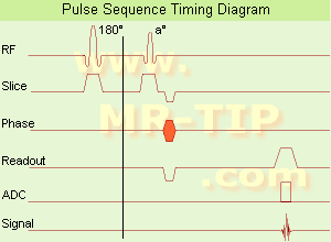
Ultrafast Gradient Echo Sequence Timing Diagram In simple ultrafast GRE imaging, TR and TE are so short, that tissues have a poor imaging signal and - more importantly - poor contrast except when contrast media enhanced (contrast enhanced angiography). Therefore, the magnetization is 'prepared' during the preparation module, most frequently by an initial 180° inversion pulse.
In the pulse sequence timing diagram, the basic ultrafast gradient echo sequence is illustrated. The 180° inversion pulse is executed one time (to the left of the vertical line), the right side represents the data collection period and is often repeated depending on the acquisition parameters.
See also Pulse Sequence Timing Diagram, there you will find a description of the components.
Ultrafast GRE sequences have a short TR,TE, a low flip angle and TR is so short that image acquisition lasts less than 1 second and typically less than 500 ms. Common TR: 3-5 msec, TE: 2 msec, and the flip angle is about 5°. Such sequences are often labeled with the prefix 'Turbo' like TurboFLASH, TurboFFE and TurboGRASS.
This allows one to center the subsequent ultrafast GRE data acquisition around the inversion time TI, where one of the tissues of interest has very little signal as its z-magnetization is passing through zero.
Unlike a standard inversion recovery (IR) sequence, all lines or a substantial segment of k-space image lines are acquired after a single inversion pulse, which can then together be considered as readout module. The readout module may use a variable flip angle approach, or the data acquisition may be divided into multiple segments (shots). The latter is useful particularly in cardiac imaging where acquiring all lines in a single segment may take too long relative to the cardiac cycle to provide adequate temporal resolution.
If multiple lines are acquired after a single pulse, the pulse sequence is a type of gradient echo echo planar imaging (EPI) pulse sequence.

See also Magnetization Prepared Rapid Gradient Echo (MPRAGE) and Turbo Field Echo (TFE).
spacer

• View the DATABASE results for 'Ultrafast Gradient Echo Sequence' (13).Open this link in a new window

MRI Resources 
Claustrophobia - Services and Supplies - Bioinformatics - Implant and Prosthesis pool - Abdominal Imaging -
 
Balanced Fast Field EchoInfoSheet: - Sequences - 
Intro, 
Overview, 
Types of, 
etc.MRI Resource Directory:
 - Sequences -
 
(bFFE) A FFE sequence using a balanced gradient waveform. A balanced sequence starts out with a RF pulse of 90° or less and the spins in the steady state. Before the next TR in the slice phase and frequency encoding, gradients are balanced so their net value is zero. Now the spins are prepared to accept the next RF pulse, and their corresponding signal can become part of the new transverse magnetization. Since the balanced gradients maintain the transverse and longitudinal magnetization, the result is, that both T1 and T2 contrast are represented in the image. This pulse sequence produces images with increased signal from fluid, along with retaining T1 weighted tissue contrast. Because this form of sequence is extremely dependent on field homogeneity, it is essential to run a shimming prior the acquisition. A fully balanced (refocused) sequence would yield higher signal, especially for tissues with long T2 relaxation times.

See Steady State Free Precession and Gradient Echo Sequence.
 
Images, Movies, Sliders:
 Cardiac Infarct Short Axis Cine bFFE 1  Open this link in a new window
    
 
spacer

• View the DATABASE results for 'Balanced Fast Field Echo' (3).Open this link in a new window

 
Further Reading:
  News & More:
T1rho-prepared balanced gradient echo for rapid 3D T1rho MRI
Monday, 1 September 2008   by www.ncbi.nlm.nih.gov    
Utility of the FIESTA Pulse Sequence in Body Oncologic Imaging: Review
June 2009   by www.ajronline.org    
Searchterm 'Magnetization' was also found in the following services: 
spacer
News  (1)  Forum  (3)  
 
Balanced SequenceForum -
related threadsInfoSheet: - Sequences - 
Intro, 
Overview, 
Types of, 
etc.
 
This family of sequences uses a balanced gradient waveform. This waveform will act on any stationary spin on resonance between 2 consecutive RF pulses and return it to the same phase it had before the gradients were applied. A balanced sequence starts out with a RF pulse of 90° or less and the spins in the steady state. Prior to the next TR in the slice encoding, the phase encoding and the frequency encoding direction, gradients are balanced so their net value is zero. Now the spins are prepared to accept the next RF pulse, and their corresponding signal can become part of the new transverse magnetization. If the balanced gradients maintain the longitudinal and transverse magnetization, the result is that both T1 and T2 contrast are represented in the image.
This pulse sequence produces images with increased signal from fluid (like T2 weighted sequences), along with retaining T1 weighted tissue contrast. Balanced sequences are particularly useful in cardiac MRI. Because this form of sequence is extremely dependent on field homogeneity, it is essential to run a shimming prior the acquisition.
Usually the gray and white matter contrast is poor, making this type of sequence unsuited for brain MRI. Modifications like ramping up and down the flip angles can increase signal to noise ratio and contrast of brain tissues (suggested under the name COSMIC - Coherent Oscillatory State acquisition for the Manipulation of Image Contrast).
These sequences include e.g. Balanced Fast Field Echo (bFFE), Balanced Turbo Field Echo (bTFE), Fast Imaging with Steady Precession (TrueFISP, sometimes short TRUFI), Completely Balanced Steady State (CBASS) and Balanced SARGE (BASG).
 
Images, Movies, Sliders:
 Cardiac Infarct Short Axis Cine Overview  Open this link in a new window
    

Courtesy of  Robert R. Edelman
 Infarct 4 Chamber Cine  Open this link in a new window
    

Courtesy of  Robert R. Edelman
 
spacer

• View the DATABASE results for 'Balanced Sequence' (5).Open this link in a new window

 
Further Reading:
  News & More:
Generic Eddy Current Compensation for Rapid Magnetic Resonance Imaging(.pdf)
   by www.switt.ch    
Magnetic resonance imaging guided musculoskeletal interventions at 0.23T: Chapter 4. Materials and methods
2002
Searchterm 'Magnetization' was also found in the following service: 
spacer
Radiology  (3) Open this link in a new window
Continuous Arterial Spin LabelingInfoSheet: - Sequences - 
Intro, 
Overview, 
Types of, 
etc.
 
(CASL) In order to take spins in a different magnetic state to that of the static tissue a train of RF pulses repeatedly saturates blood water spins flowing through the neck. The saturated (or 'labeled') spins flow into the brain and, assuming water is a freely diffusible tracer, exchange completely with brain tissue water, thus reducing the overall tissue magnetization. A steady state progresses where the regional magnetization in the brain is directly related to cerebral blood flow.
spacer
 
Further Reading:
  Basics:
Measuring Cerebral Blood Flow Using Magnetic Resonance Imaging Techniques
1999   by www.stanford.edu    
  News & More:
Non-invasive MRI technique distinguishes between Alzheimer's and frontotemporal dementia
Saturday, 18 June 2005   by www.eurekalert.org    
MRI Resources 
Examinations - MRI Physics - Cochlear Implant - Lung Imaging - MRI Accidents - Absorption and Emission
 
previous      36 - 40 (of 127)     next
Result Pages : [1 2 3]  [4 5 6 7 8 9 10 11 12 13 14 15 16 17 18 19 20 ... ]
 Random Page
 
Share This Page
FacebookTwitterLinkedIn

MR-TIP    
Community   
User
Pass
Forgot your UserID/Password ?    


How AI will impact MRI :
only diagnostics 
saving time 
reducing cost 
makes planning obsolete 
reduce human knowledge 
not at all 

Look
      Ups


MR-TIP.com uses cookies! By browsing MR-TIP.com, you agree to our use of cookies.

Magnetic Resonance - Technology Information Portal
Member of SoftWays' Medical Imaging Group - MR-TIP • Radiology-TIP • Medical-Ultrasound-Imaging • 
Copyright © 2003 - 2024 SoftWays. All rights reserved. [ 9 May 2024]
Terms of Use | Privacy Policy | Advertising
 [last update: 2024-02-26 03:41:00]