|
Magnetic
Resonance -
Technology
Information
Portal |
Welcome to MRI Technology • • |
|
|
 | Info
Sheets |
| | | | | | | | | | | | | | | | | | | | | | | | |
 | Out-
side |
| | | | |
|
| | | | | |
| Result: Searchterm 'Meter'
found in 42 messages |
| Result Pages: 1 2 [3] 4 5 6 7 8 9 |
More Results:  Database (127) News Service (31) Resources (36) Database (127) News Service (31) Resources (36) |
|
|
Edmund Kwok
Tue. 20 Oct.15,
20:34
[Start of:
'Artifact in Abdomen LAVA'
0 Reply]
 Category: Category:
Artifacts
|
| Artifact in Abdomen LAVA |
This artifact appears on every slice of an abdomen LAVA scan, and it looks like puzzle pieces. The artifact was gone in a re-scan using similar parameters. Does any one has an idea what might have caused it? Thanks.
|
| |  Reply to this thread Reply to this thread
(login or register first) | |
marc jupin
Thu. 26 Mar.15,
09:03
[Start of:
'receiver gain in siemens trio'
0 Reply]
Category:
Sequences and Imaging Parameters
|
| receiver gain in siemens trio |
Hi, we measured a phantom with two receiver gain option: LOW and HIGH (available from parameters).
We would like to calculate the difference in signal intensity, but unfortunately the rda files headers do not contain any information about the receiver gain value.
Does anyone have a clue how I could find the receiver gain value?
Thanks.
|
| | Reply to this thread
(login or register first) | |
Bob Smtih
Thu. 30 Oct.14,
18:41
[Start of:
'What is this white "cloud" on the right side of this MRI?'
0 Reply]
Category:
General
|
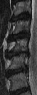
| What is this white "cloud" on the right side of this MRI? |
The following was noted:c2/c3 normal c3/c4 Very minimal anterior ostepphytic ridging is present. There is no spinal stenosis or neural foraminal narrowing. C4/c5 Very minimal anterior ostepphytic ridging is noted. There is no spinal stenosis or neural foraminal narrowing. c5/c6 There is a circumferential disc osteophyte complex present. There is mild central spinal stenosis. The AP diameter of the spinal canal is narrowed to 9mm. The dorsal and ventral CSF spaces remain patent. There is mild to moderate neural foraminal narrowing. c6/c7 a circumferential disc osteophyte complex present. There is mild central spinal stenosis. AP diameter of the spinal canal is narrowed to 9mm. The dorsal and ventral CSF spaces remain patent. There is moderate to severe neural foraminal narrowing. c7/t1 Normal. AP diameter of the spinal canal is 12 mm My question is C5 looks like it is chewed up; weird shading. Does it appear to be more damaged than the report states? Also there seems to be a white "cloud" that goes into the left side of multiple disks. What is the white "cloud"? c3 through c7 are in the image
 MRI SAG T2 MRI SAG T2
|
| | Reply to this thread
(login or register first) | |
Niels Janssen
Tue. 6 May.14,
11:24
[Start of:
'GE signa excite 3T - clustered volume acquisition'
0 Reply]
Category:
Funktional MRI
|
| GE signa excite 3T - clustered volume acquisition |
I am wondering if anyone has experience with so called 'clustered volume acquisition' on a GE signa excite 3T. Clustered volume acquisition (or sparse acquisition) means that you acquire images within the first 1000 ms of a 2000 ms TR, for example (Edmister et al., 1999, Human Brain Mapping). This method is useful because the machine will stop producing scanning noise in the final 1000 ms of the TR, which can then be used for recording speech inside the scanner (in fmri applications).
I am asking whether 1) this sequence can be programmed from the parameters menu on the scanner console, or 2) whether it requires compilation of a new pulse sequence.
|
| | Reply to this thread
(login or register first) | |
Reader Mail
Fri. 26 Jul.13,
16:51
[Reply (1 of 2) to:
'STIR and l-spine'
started by: 'shruti soni'
on Wed. 27 Feb.13]
Category:
Sequences and Imaging Parameters
|
| STIR and l-spine |
I know from working on various coils and testing the images will vary with synthetic (phantoms) vs actual patient. Main item in coil repair is it scans correct w/a human. And if when you test the coil per Philips given guidelines and set parameters for testing SNR, etc. the coil may be just fine. 2nd the synthetic phantom your using is not compatible with that coil or in the system - found this out with 1.5T phantoms don't work on 0.2T coils.
|
| | View the whole thread | |
| |
| | Result Pages : 1 2 [3] 4 5 6 7 8 9 | |
|
| |
 | Look
Ups |
| |
|
MR-TIP.com uses cookies! By browsing MR-TIP.com, you agree to our use of cookies. | | | [last update: 2024-02-26 03:41:00] |
|
|


