|
Magnetic
Resonance -
Technology
Information
Portal |
Welcome to MRI Technology • • |
|
|
 | Info
Sheets |
| | | | | | | | | | | | | | | | | | | | | | | | |
 | Out-
side |
| | | | |
|
| | | | | |
| Result: Searchterm 'Spin'
found in 59 messages |
| Result Pages: 1 2 3 4 [5] 6 7 8 9 10 11 12 |
More Results:  Database (332) News Service (64) Resources (27) Database (332) News Service (64) Resources (27) |
|
|
William Richardson
Tue. 20 Jun.17,
12:33
[Start of:
'MRI 1w/kg sequences Siemens Avanto FIT'
0 Reply]
 Category: Category:
Sequences and Imaging Parameters
|
| MRI 1w/kg sequences Siemens Avanto FIT |
I have recently been asked to build protocols for Spines and extremities that are 1 watt per kilogram SAR. In speaking with Applications at Siemens they inform me that I can only change certian parameters to help lower SAR but it will all be dependent upon each patient and which sequences are running. I need to present to our MRI QA commitiee if we can or if we cannot do 1w/kg SAR as we are determining if we can scan cochlear implants? Does anyone have any ideas or solutions?
Thank You
Will R.
|
| |  Reply to this thread Reply to this thread
(login or register first) | |
John Smith
Wed. 11 Nov.15,
22:14
[Start of:
'Faster pulse sequences'
0 Reply]
Category:
General
|
| Faster pulse sequences |
Hi,
I have been learning about faster MRI sequences and have two questions
1) With "Fast (Turbo) gradient echo", in which we apply a spoiler gradient, do we not eventually end up with no longitudinal magnetization because TR is always shorter than T1? Hence shouldn't we eventually get no signal at all?
2) in SSFP (Steady-state free precession) we can apply an RF pulse of 90 degrees (in which T1>>T2) to get heart-blood contrast. How is this any different to a standard spin-echo sequence in terms of timing?
Thank you
|
| | Reply to this thread
(login or register first) | |
Bob Smtih
Thu. 30 Oct.14,
18:41
[Start of:
'What is this white "cloud" on the right side of this MRI?'
0 Reply]
Category:
General
|
| What is this white "cloud" on the right side of this MRI? |
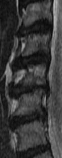
The following was noted:c2/c3 normal c3/c4 Very minimal anterior ostepphytic ridging is present. There is no spinal stenosis or neural foraminal narrowing. C4/c5 Very minimal anterior ostepphytic ridging is noted. There is no spinal stenosis or neural foraminal narrowing. c5/c6 There is a circumferential disc osteophyte complex present. There is mild central spinal stenosis. The AP diameter of the spinal canal is narrowed to 9mm. The dorsal and ventral CSF spaces remain patent. There is mild to moderate neural foraminal narrowing. c6/c7 a circumferential disc osteophyte complex present. There is mild central spinal stenosis. AP diameter of the spinal canal is narrowed to 9mm. The dorsal and ventral CSF spaces remain patent. There is moderate to severe neural foraminal narrowing. c7/t1 Normal. AP diameter of the spinal canal is 12 mm My question is C5 looks like it is chewed up; weird shading. Does it appear to be more damaged than the report states? Also there seems to be a white "cloud" that goes into the left side of multiple disks. What is the white "cloud"? c3 through c7 are in the image
 MRI SAG T2 MRI SAG T2
|
| | Reply to this thread
(login or register first) | |
Reader Mail
Wed. 29 Oct.14,
20:38
[Start of:
'Vibrant Sound Bridge and MRI'
0 Reply]
Category:
Devices, Scanner, Machines
|
| Vibrant Sound Bridge and MRI |
Hello everyone,
I have a Vibrant Soundbridge implanted on both sides of my skull, which contain a magnet. I use them due to severe hearing impairment.
Now the question is, I was told that MRI was prohibited for patients with a Vibrant Soundbridge.
Could anyone suggest an alternative to this problem please? Is there another equipment allowing me to have the conclusions normally obtained from MRI? My problem is an accident to my spinal cord, which requires further investigation using an MRI.
I would appreciate any help!
Thanks a lot
|
| | Reply to this thread
(login or register first) | |
Belinda Williams
Mon. 21 Jul.14,
21:44
[Start of:
'?'
1 Reply]
Category:
Coils
|
| ? |
I work on GE excite open speed. On lumbar spine t2 axials, it appears like the coil looses signal and makes image dark and grainy. The coil gives good signal on the other sequences and tests good when engineer has tested it. The sag t2 is bright like it should but ax is dark. Any ideas why?
|
| | View the whole thread | Reply to this thread
(login or register first) |
| |
| | Result Pages : 1 2 3 4 [5] 6 7 8 9 10 11 12 | |
|
| |
 | Look
Ups |
| |
|
MR-TIP.com uses cookies! By browsing MR-TIP.com, you agree to our use of cookies. | | | [last update: 2025-04-05 02:38:00] |
|
|


