|
Magnetic
Resonance -
Technology
Information
Portal |
Welcome to MRI Technology • • |
|
|
 | Info
Sheets |
| | | | | | | | | | | | | | | | | | | | | | | | |
 | Out-
side |
| | | | |
|
| | | | | |
| Result: Searchterm 'Spin'
found in 59 messages |
| Result Pages: 1 2 3 4 5 6 7 8 9 [10] 11 12 |
More Results:  Database (332) Database (332)  News Service (64) News Service (64)  Resources (27) Resources (27) |
|
|
Reader Mail
Sun. 27 Jan.08,
08:47
[Start of:
'Plz Answer this ... Contrast MRI of Brain'
1 Reply]

 Category: Category:
Applications and Examinations
|
| Plz Answer this ... Contrast MRI of Brain |
Pre and Post contrast mri of the brain was performed in multiple planes using T1 & T2 W spin-echo sequence.
There is small ring enhancing lesion in the left occipitoparietal lobe which measures 1cm in diameter.It reveals isointense periphery on T1 & T2W images with hyperintense core on T2W images. On T1W images the core appears hypointense . A tiny mural nodule is seen within the lesion. focal perilesional edema is seen appearing hyperintense on FLAIR and T2W images.
The brainstem & cerebellum are normal.
The ventricular system is normal.
No abnormal meningeal enhancement is seen.
Intracranial vessels display normal flow void.
What needs to be done?? How serious the problem is??
|
|  View the whole thread View the whole thread |  Reply to this thread Reply to this thread
(login or register first) | |
miri leh
Wed. 9 Jan.08,
09:56
[Start of:
'scan question from a layperson'
2 Replies]

 Category: Category:
Applications and Examinations
|
| scan question from a layperson |
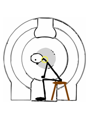
When positioned as in picture (neck bent halfway forward) in GE's Signa SP/i 0.5T, will the scan include a view of the cervical spine from the back?
Will the scan be able to fully view the vertebras (from the back of the vertebras) that are tilted due to the neck bending forward?
(why the back: positional scoliosis suspected. suspected cause: asymmetric work of the suboccipital muscles, due to severed muscle connection/s)
 MRIquestion MRIquestion


|
|  View the whole thread View the whole thread |  Reply to this thread Reply to this thread
(login or register first) | |
Reader Mail
Wed. 12 Dec.07,
10:07
[Reply (1 of 2) to:
'double ir physic'
started by: 'soontorn siriserussa'
on Sun. 2 Dec.07]

 Category: Category:
Sequences and Imaging Parameters
|
| double ir physic |
Different types of double inversion recovery (DIR, 2IR) sequences are used to improve the suppression of blood signal (black blood technique) or to null the signals from two different tissue types (e.g. white matter and cerebrospinal fluid).
The black blood technique (used in cardiovascular MRI) works with two inversion pulses, where the first pulse is nonselective and the second pulse is slice-selective. TI is set to a value at which the signal of the recovering inverted blood is zero (http://www.mr-tip.com/serv1.php?type=db1&dbs=Double%20Inversion).
The second technique (also named gray matter only) is used in brain imaging to improve the detection of lesions, for example in the diagnostic of multiple sclerosis. Two 180° pulses with different TI are used to suppress two different types of tissue simultaneously.
Hope this helps
|
|  View the whole thread View the whole thread | | |
Jenny Jordan
Sat. 17 Nov.07,
20:11
[Reply (2 of 4) to:
'Haste and Rare sequences'
started by: 'Elena sussi'
on Tue. 13 Nov.07]

 Category: Category:
Sequences and Imaging Parameters
|
| Haste and Rare sequences |
Hi Elena,
you can find the different names for the same sequences and options used by manufacturers at: http://www.mr-tip.com/serv1.php?type=cam .
The manufacturers have in principle similar sequences with small differences. So, as you mentioned you can take "sequences as HASTE" or "sequences type RARE".
However, you can lead back these sequences also to the fundamental sequence type FSE ( fast spin echo: http://www.mr-tip.com/serv1.php?type=seq&sub=12 ). Fast sequences as HASTE ( http://www.mr-tip.com/serv1.php?type=db1&dbs=haste ) are especially useful in cases of movement caused by their single shot technique: http://www.mr-tip.com/serv1.php?type=db1&dbs=Single%20Shot%20Technique .
Hope this helps
|
|  View the whole thread View the whole thread | | |
andri anto
Fri. 25 May.07,
06:39
[Reply (1 of 2) to:
'What´s this in L3?'
started by: 'Rob van den Dobbelsteen'
on Sat. 10 Mar.07]

 Category: Category:
Applications and Examinations
|
| What´s this in L3? |
You just give t2 sagital WI, that not enough for determined what the problem in L3 spine, better you asked to radiologist and show all the MRI picture. Cause i saw there lession in corpus L3 and bulging in discus L3-l4 (HNP). To know more about something in Corpus L3 you must have all sequence in MRI Lumbal exam.
Andri
|
|  View the whole thread View the whole thread | |
| |
| | Result Pages : 1 2 3 4 5 6 7 8 9 [10] 11 12 | |
|
| |
 | Look
Ups |
| |
|
MR-TIP.com uses cookies! By browsing MR-TIP.com, you agree to our use of cookies. | | | [last update: 2025-04-05 02:38:00] |
|
|