|
Magnetic
Resonance -
Technology
Information
Portal |
Welcome to MRI Technology • • |
|
|
 | Info
Sheets |
| | | | | | | | | | | | | | | | | | | | | | | | |
 | Out-
side |
| | | | |
|
| | | | | |
| Result: Searchterm 'Slice'
found in 36 messages |
| Result Pages: 1 2 3 [4] 5 6 7 8 |
More Results:  Database (177) News Service (6) Resources (5) Database (177) News Service (6) Resources (5) |
|
|
elena giovanelli
Thu. 24 Oct.13,
09:22
[Start of:
'mr urography'
1 Reply]
 Category: Category:
Applications and Examinations
|
| mr urography |
hello everyone.I need help with mr urography. how do you plan graphically the sequence MRCP for the study of the ureters? I mean if you considered a kidney at a time or both in the fov, how many thick slice you need. etc.
I thanks a lot
|
|  View the whole thread View the whole thread | Reply to this thread
(login or register first) | |
Brent Johnson
Wed. 21 Aug.13,
19:38
[Start of:
'Hitachi Airis II question'
0 Reply]
Category:
Sequences and Imaging Parameters
|
| Hitachi Airis II question |
I have a question for a Hitachi Airis II .3T non-upgraded gradients
On T2 FSE sequences especially on thumb sagittal slices. Image quality very poor, grainy. What is ideal bandwidth and TE settings for improving image quality? Also is positioning and angle of slice a factor in image quality. The closer the anatomy is to edge of coil, does that effect image quality.
Thanks Brent
|
| | Reply to this thread
(login or register first) | |
Abdul Sireis
Wed. 13 Jun.12,
13:20
[Start of:
'time point'
0 Reply]
Category:
Basics and Physics
|
| time point |
Hello,
I read in the references about time point but i'm not sure if it is total scan which contains many slices or that each time point is a slice itself. Can anyone here clarify something about this? Thanks
|
| | Reply to this thread
(login or register first) | |
Steven Ford
Mon. 23 Apr.12,
19:58
[Reply (2 of 3) to:
'intracranial cavity volume'
started by: 'adam wootton'
on Fri. 13 Apr.12]
Category:
Applications and Examinations
|
| intracranial cavity volume |
Osiris can do this, but it's on a slice by slice basis the last time I tried it, which is quite tedious. And it's not FDA approved for that application, I believe.
But GE probably has a tool you can use for that; why don't you ask your GE rep?
Steven Ford
Professional Imaging Services, Inc.
|
| | View the whole thread | | |
Reader Mail
Tue. 26 Jul.11,
14:05
[Start of:
'MR750: ASSET Artifact or 32 Channel coil?'
1 Reply]
Category:
Funktional MRI
|
| MR750: ASSET Artifact or 32 Channel coil? |
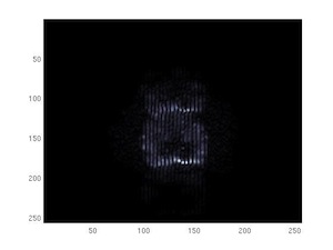
Hi,
We are performing fMRI on a GE MR750 Discovery.
As you can see we get artifacts when performing DTI and fMRI.
It looks like herringbones or spikes artifacts. But it appears only at the very top slices where there is generally no more brain.
One possible explanation is that ASSET will produce the artifact where there is no signal.
Another explanation would be that some element of the 32 channel coil is going mad.
Has anybody experienced the same artifact?
Thank you.
 DTI artifact DTI artifact
|
| | View the whole thread | Reply to this thread
(login or register first) |
| |
| | Result Pages : 1 2 3 [4] 5 6 7 8 | |
|
| |
 | Look
Ups |
| |
|
MR-TIP.com uses cookies! By browsing MR-TIP.com, you agree to our use of cookies. | | | [last update: 2024-02-26 03:41:00] |
|
|


