|
Magnetic
Resonance -
Technology
Information
Portal |
Welcome to MRI Technology • • |
|
|
 | Info
Sheets |
| | | | | | | | | | | | | | | | | | | | | | | | |
 | Out-
side |
| | | | |
|
| | | | | |
| Result: Searchterm 'Slice'
found in 36 messages |
| Result Pages: 1 2 [3] 4 5 6 7 8 |
More Results:  Database (177) News Service (6) Resources (5) Database (177) News Service (6) Resources (5) |
|
|
Haider Ali
Thu. 15 Sep.16,
02:26
[Start of:
'Toshiba Vantage Titan 3T'
0 Reply]
 Category: Category:
Equipment
|
| Toshiba Vantage Titan 3T |
While planning vertebral discs for axial cuts is there any way to align the slices perpendicularly on coronal plane? Currently I have to manually align them on the vertebral column which is tedious and sometimes erroneous.
I have worked on Siemens Avanto and there is option to select the slice to align perpendicular so that I only have to pay attention to one plane while in Toshiba the task is a bit more complicated. Though there is an option of aligning all slices perpendicularly but it does not work or may be I am doing something wrong.
|
| |  Reply to this thread Reply to this thread
(login or register first) | |
Marlee Smith
Sat. 23 Jan.16,
00:45
[Start of:
'Brain MRI Help'
0 Reply]
Category:
General
|
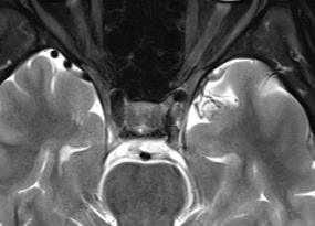
| Brain MRI Help |
Can someone please help me interpret this horizontal MRI T2 slice? The images are of just behind my eyes. I don't think those black dots are meant to be there... I know that foramen make a black dot but I don't think that there are any foramen where the dots are.
|
| | Reply to this thread
(login or register first) | |
Edmund Kwok
Tue. 20 Oct.15,
20:34
[Start of:
'Artifact in Abdomen LAVA'
0 Reply]
Category:
Artifacts
|
| Artifact in Abdomen LAVA |
This artifact appears on every slice of an abdomen LAVA scan, and it looks like puzzle pieces. The artifact was gone in a re-scan using similar parameters. Does any one has an idea what might have caused it? Thanks.
|
| | Reply to this thread
(login or register first) | |
dimitris priovolos
Sat. 21 Dec.13,
10:14
[Reply (1 of 2) to:
'mr urography'
started by: 'elena giovanelli'
on Thu. 24 Oct.13]
Category:
Applications and Examinations
|
| mr urography |
I think it's better to plan both ureters, put the pack of slices exactly coronal on an axial abdomen slice including both kidneys, take care on a sagital slice to give an angle parallel to spine but including the bladder and the space behind it, use foldover direction (siemens) or Rfov 1 (GE), fov 400-450, sl thick 0.80-1mm, open the 3d slice pack as much as needed, use navigator-pace (siemens) or triggering (GE).
A preparation of diet is needed some times, additional sequences (haste, fiesta, vibe+iv or lava+iv contr) are necessary.
|
| | View the whole thread | | |
dimitris priovolos
Sat. 21 Dec.13,
09:40
[Reply (1 of 2) to:
'Can a pancoast tumor show up on a cervical spine MRI?'
started by: 'Lucy Lane'
on Wed. 28 Aug.13]
Category:
Applications and Examinations
|
| Can a pancoast tumor show up on a cervical spine MRI? |
I think yes,sagital plan can give you a sign on left or right side (T1W, TW2), then on coronal plan and with thicker slices you can localize it (like doing a brachio plexus exam).
|
| | View the whole thread | |
| |
| | Result Pages : 1 2 [3] 4 5 6 7 8 | |
|
| |
 | Look
Ups |
| |
|
MR-TIP.com uses cookies! By browsing MR-TIP.com, you agree to our use of cookies. | | | [last update: 2024-02-26 03:41:00] |
|
|


