|
Magnetic
Resonance -
Technology
Information
Portal |
Welcome to MRI Technology • • |
|
|
 | Info
Sheets |
| | | | | | | | | | | | | | | | | | | | | | | | |
 | Out-
side |
| | | | |
|
| | | | | |
| Result: Searchterm 'Axial'
found in 17 messages |
| Result Pages: 1 [2] 3 4 |
More Results:  Database (21) News Service (2) Resources (2) Database (21) News Service (2) Resources (2) |
|
|
dimitris priovolos
Sat. 21 Dec.13,
10:14
[Reply (1 of 2) to:
'mr urography'
started by: 'elena giovanelli'
on Thu. 24 Oct.13]
 Category: Category:
Applications and Examinations
|
| mr urography |
I think it's better to plan both ureters, put the pack of slices exactly coronal on an axial abdomen slice including both kidneys, take care on a sagital slice to give an angle parallel to spine but including the bladder and the space behind it, use foldover direction (siemens) or Rfov 1 (GE), fov 400-450, sl thick 0.80-1mm, open the 3d slice pack as much as needed, use navigator-pace (siemens) or triggering (GE).
A preparation of diet is needed some times, additional sequences (haste, fiesta, vibe+iv or lava+iv contr) are necessary.
|
|  View the whole thread View the whole thread | | |
Karen Lesley
Thu. 26 Jan.12,
15:55
[Reply (4 of 7) to:
'Imaging optic neuritis'
started by: 'Karen Lesley'
on Wed. 18 Jan.12]
Category:
General
|
| Imaging optic neuritis |
Thanks Anna :-)
I've recommended thin coronal STIR (the "axial" in my original post was a mistake - I do know better, promise!) and FLAIR.
The ON is longstanding, so may not show with gadolinium, but worth a try if the budget will stretch so will add that too.
Thanks again.
|
| | View the whole thread | | |
Karen Lesley
Wed. 18 Jan.12,
19:11
[Start of:
'Imaging optic neuritis'
6 Replies]
Category:
General
|
| Imaging optic neuritis |
Hope someone can help!
Is there a better way to image optic neuritis than thin axial FLAIR?
Thanks in advance!
|
| | View the whole thread | Reply to this thread
(login or register first) | |
Mel Chang
Thu. 21 Oct.10,
19:26
[Reply (1 of 2) to:
'cervicla axial image t2 and t2*'
started by: 'kim jk'
on Thu. 23 Sep.10]
Category:
Applications and Examinations
|
| cervicla axial image t2 and t2* |
T2* is fine to scan the intervertebral discs. T2 is the better choice to show small spinal cord lesions like MS plaques. The echo time of a T2* sequence may be not long enough to give a good differentiation between the pathology and surrounding tissue.rnDifferent 'Multi Echo Data Image Combination' (MEDIC) techniques have been developed to enhance contrast and pathology detection, if available on the scanner this type of sequence is maybe also a good choice, but due to longer scan times and artifact problems it is best for axial slices.rn
|
| | View the whole thread | | |
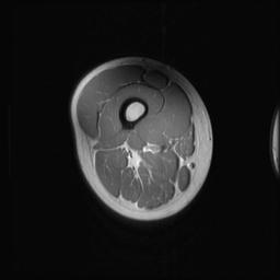
Travis Conley
Thu. 21 Oct.10,
17:22
[Start of:
'Muscle shading in 3T Images'
2 Replies]
Category:
Applications and Examinations
|
| Muscle shading in 3T Images |
We are using a 3T GE scanner to image axial slices of human thigh muscle for the purpose of quantifying muscle volume. rnrnProblem: Our images have been presenting some shading at the top and bottom of each slice. We have tried everything we can think of to get rid of this shading but have not been sucessful. rnrnI have attached a representative image and would love some feel back. rnrnThanks so much for attempting to help us out.
 Thigh Axial Slice Thigh Axial Slice
|
| | View the whole thread | Reply to this thread
(login or register first) |
| |
| | Result Pages : 1 [2] 3 4 | |
|
| |
 | Look
Ups |
| |
|
MR-TIP.com uses cookies! By browsing MR-TIP.com, you agree to our use of cookies. | | | [last update: 2024-02-26 03:41:00] |
|
|


