Magnetic Resonance - Technology Information Portal Welcome to MRI Technology
Info
  Sheets

Out-
      side
 

How AI will impact MRI :
only diagnostics 
saving time 
reducing cost 
makes planning obsolete 
reduce human knowledge 
not at all 


MRI Artifacts
  
Ultrasound Imaging Artifacts Open this link in a new window
Susceptibility Artifact 
Quick Overview
Artifact Information
DESCRIPTION
Signal dropout, bright spots, spatial distortion
REASON
HELP
Remove the metal, do not take a gradient echo sequence, take a short echo time
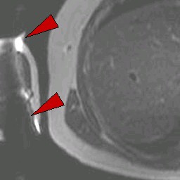
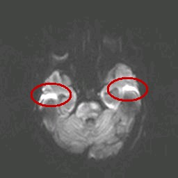
Materials with magnetic susceptibility cause this artifact. There are in general three kinds of materials with magnetic susceptibility: ferromagnetic materials (iron, nickel etc.) with a strong influence and paramagnetic/diamagnetic (aluminium, platinum etc./gold, water, most organic compounds etc.) materials with a minimal/non influence on magnetic fields. In MRI, susceptibility artifacts are caused for example by medical devices in or near the magnetic field or by implants of the patient. These materials with magnetic susceptibility distort the linear magnetic field gradients, which results in bright areas (misregistered signals) and dark areas (no signal) nearby the magnetic material.
mri safety guidance
Image Guidance
Use a spin echo or a fast spin echo sequence, because gradient echo sequences are more sensitve to susceptibility artifacts. A high bandwidth (small water fat shift) and a short echo time help also to reduce this artifact.
In some cases it is even beneficial to use a gradient echo sequence, e.g. a cavernom contains some iron-rich haemosiderin, which also causes a signal void on gradient echo sequences and for this purpose increases the diagnostic image quality.

• View the DATABASE results for 'Susceptibility Artifact' (8).Open this link in a new window

 
Further Reading:
  Basics:
MRI Artifact Gallery
   by chickscope.beckman.uiuc.edu    
Susceptibility Artifacts
   by www.mritutor.org    
  News & More:
Metal Artefact Reduction
Thursday, 9 June 2011   by www.revisemri.com    
Ultrashort echo time (UTE) MRI of the spine in thalassaemia
February 2004   by bjr.birjournals.org    
Case Study 1 :
This artifact is caused by a metallic application on the sleeve.
 
Case Study 2 :
This artifact is commonly visible in diffusion imaging at the interface between different tissues at the cranial basic. Bright and dark areas show the spatial distortion.
 
Similar Artifacts:
 • Metal Artifact
 • Field Inhomogeneity Artifact
 • Moire Fringes (Artifact)
  Truncation Artifact top
 
Share This Page
FacebookTwitterLinkedIn

MR-TIP    
Community   
User
Pass
Forgot your UserID/Password ?    


Look
      Ups


MR-TIP.com uses cookies! By browsing MR-TIP.com, you agree to our use of cookies.

Magnetic Resonance - Technology Information Portal
Member of SoftWays' Medical Imaging Group - MR-TIP • Radiology-TIP • Medical-Ultrasound-Imaging • 
Copyright © 2003 - 2024 SoftWays. All rights reserved. [ 25 April 2024]
Terms of Use | Privacy Policy | Advertising
 [last update: 2024-02-26 03:41:00]