Magnetic Resonance - Technology Information Portal Welcome to MRI Technology
Info
  Sheets

Out-
      side
 

MRI is trending to low field magnets :
reduced costs will lead to this change 
AI will close the gap to high field 
only in remote areas 
is only temporary 
never 


MRI Sliders
  Sliders Overview
 MRI - Anatomic Imaging of the Ankle 2  
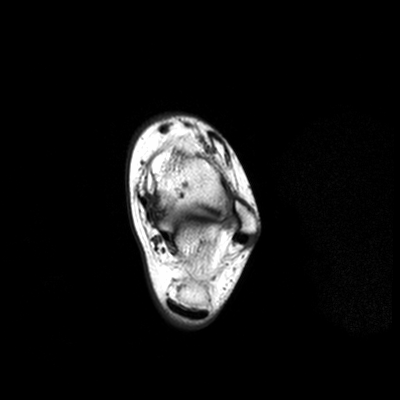
The interactive T1 weighted image series shows the anatomy of the ankle in the transverse plane from the calcaneum (sole of the foot, slice 1) to the distal fibula and tibia. T1 weighted transverse MR images provides good visualization of the bones, surrounding ligaments, muscles, fat and the achilles tendon.
Slice:   (of 24)

 
Related Images
 • MRI Orbita T1
 • Knee MRI Sagittal T1 001
 • Knee MRI Sagittal T1 002
 • Knee MRI Sagittal T1 003
 • Knee MRI Sagittal T1 004
 • Sagittal Knee MRI Images T1 Weighted
Relevant Database Entries
 • T1 Weighted Image
 • T1 Time
 • Imaging of the Extremities


  Next Slider: MRI - Anatomic Imaging of the Ankle 3 top
Electricity is actually made up of extremely tiny particles called electrons, that you cannot see with the naked eye unless you have been drinking.
- Dave Barry
 
Share This Page
FacebookTwitterLinkedIn

MR-TIP    
Community   
User
Pass
Forgot your UserID/Password ?    


Look
      Ups


MR-TIP.com uses cookies! By browsing MR-TIP.com, you agree to our use of cookies.

Magnetic Resonance - Technology Information Portal
Member of SoftWays' Medical Imaging Group - MR-TIP • Radiology-TIP • Medical-Ultrasound-Imaging • 
Copyright © 2003 - 2024 SoftWays. All rights reserved. [ 24 April 2024]
Terms of Use | Privacy Policy | Advertising
 [last update: 2024-02-26 03:41:00]