|
MRI - Anatomic Imaging of the Ankle 1 |
|
 |
|
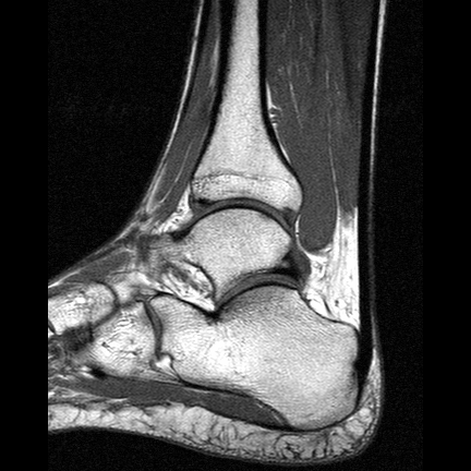
| This interactive T1 weighted image series shows the anatomy of the ankle, seen in a sagittal view with high spatial resolution and thin slices. T1 weightings produce high-intensity signals of fat and bone marrow and low-intensity signals of muscles, water and collagen tissues.
The talocrural joint is formed by the distal ends of the fibula (lateral or outside portion of the ankle, the first slices) the tibia (medial or inside portion of the ankle) and the talus. The medial and lateral collateral ligaments prevent abduction and adduction. The subtalar joint consists of the talus on top and calcaneus on the bottom. | |
| |
|
|
| |
| |
| Related Images
• MRI Orbita T1
• Knee MRI Sagittal T1 001
• Knee MRI Sagittal T1 002
• Knee MRI Sagittal T1 003
• Knee MRI Sagittal T1 004
• Sagittal Knee MRI Images T1 Weighted |
|
| Relevant Database Entries
• MRI
• Shoulder MRI
• MRI of the Brain
• Imaging of the Extremities
• Knee MRI
• MRI Scan
• T1 Time |
|
|
| |