Magnetic Resonance - Technology Information Portal Welcome to MRI Technology
Info
  Sheets

Out-
      side
 

Personalized protocols (age, gender, body habitus, etc.) lead to :
more automated planning 
improved patient comfort 
better diagnostics 
optimized image quality 
nothing 


MRI Sequences


ball_redInversion Recovery Sequence 
Inversion Recovery Sequence Timing Diagram (IR) The inversion recovery pulse sequence produces signals, which represent the longitudinal magnetization existing after the application of a 180° radio frequency pulse that rotates the magnetization Mz into the negative plane. After an inversion time (TI - time between the starting 180° pulse and the following 90° pulse), a further 90° RF pulse tilts some or all of the z-magnetization into the xy-plane, where the signal is usually rephased with a 180° pulse as in the spin echo sequence. During the initial time period, various tissues relax with their intrinsic T1 relaxation time.
In the pulse sequence timing diagram, the basic inversion recovery sequence is illustrated. The 180° inversion pulse is attached prior to the 90° excitation pulse of a spin echo acquisition. See also the Pulse Sequence Timing Diagram. There you will find a description of the components.
The inversion recovery sequence has the advantage, that it can provide very strong contrast between tissues having different T1 relaxation times or to suppress tissues like fluid or fat. But the disadvantage is, that the additional inversion radio frequency RF pulse makes this sequence less time efficient than the other pulse sequences.

Contrast values:
PD weighted: TE: 10-20 ms, TR: 2000 ms, TI: 1800 ms
T1 weighted: TE: 10-20 ms, TR: 2000 ms, TI: 400-800 ms
T2 weighted: TE: 70 ms, TR: 2000 ms, TI: 400-800 ms

See also Inversion Recovery, Short T1 Inversion Recovery, Fluid Attenuation Inversion Recovery, and Acronyms for 'Inversion Recovery Sequence' from different manufacturers.

• View the DATABASE results for 'Inversion Recovery Sequence' (8).Open this link in a new window

 
Further Reading:
  Basics:
The equation for a repeated inversion recovery sequence
Contrast mechanisms in magnetic resonance imaging
2004   by www.iop.org    
  News & More:
FLAIR Vascular Hyperintensity: An Important MRI Marker in Patients with Transient Ischemic Attack
Thursday, 14 July 2022   by www.dovepress.com    
Flow Sensitive Alternating Inversion Recovery 
(FAIR) In this sequence 2 inversion recovery images are acquired, one with a nonselective and the other with a slice selective inversion pulse. The z-magnetization in the first sequence is independent of flow. Inflowing spins give z-magnetization from second pulse. A major signal loss in FAIR is the T1 relaxation of tagged blood in transit to the imaging slice. Sharper edges of the inversion pulse give narrow spacing between the inversion edge and the 1st slice because reduced transit time gives lower T1 relaxation induced signal loss. The difference of the images in a consequence contains information proportional to flow (blood partition coefficient). Standard adiabatic inversion RF pulse does not have good slice-profile, because of power/SAR limitation. A c-shaped frequency offset corrected inversion (FOCI) RF pulse can help to increase the signal.
Perfusion imaging, e.g. myocardial, using tissue water as endogenous contrast is suggested.
Fluid Attenuation Inversion Recovery 
(FLAIR) Fluid attenuation inversion recovery is a special inversion recovery sequence with long TI to remove the effects of fluid from the resulting images. The TI time of the FLAIR pulse sequence is adjusted to the relaxation time of the component that should be suppressed. For fluid suppression the inversion time (long TI) is set to the zero crossing point of fluid, resulting in the signal being 'erased'.
Lesions that are normally covered by bright fluid signals using conventional T2 contrast are made visible by the dark fluid technique FLAIR is an important technique for the differentiation of brain and spine lesions.

See also Inversion Recovery.

• View the DATABASE results for 'Fluid Attenuation Inversion Recovery' (5).Open this link in a new window

 
Further Reading:
  Basics:
Newer Sequences for Spinal MR Imaging: Smorgasbord or Succotash of Acronyms?
   by www.ajnr.org    
  News & More:
What MRI-Derived Data and Other Factors Reveal About White Matter Hyperintensity in Former Football Players
Saturday, 23 December 2023   by www.diagnosticimaging.com    
FLAIR Vascular Hyperintensity: An Important MRI Marker in Patients with Transient Ischemic Attack
Thursday, 14 July 2022   by www.dovepress.com    
Early Identification of Ischemic Stroke With DWI-FLAIR Mismatch
Wednesday, 5 January 2011   by www.doctorslounge.com    
Inversion Recovery Spin Echo 
(IRSE) Form of inversion recovery imaging in which the signal is detected as a spin echo. For TE short compared to the T2 relaxation time, there will be only a small effect of T2 differences on image intensities; for longer TE's, the effect of T2 may be significant.
 
Further Reading:
  Basics:
Magnetic resonance imaging
   by www.scholarpedia.org    
Short T1 Inversion Recovery 
(STIR) Also called Short Tau (t) (inversion time) Inversion Recovery. STIR is a fat suppression technique with an inversion time t = T1 ln2 where the signal of fat is zero (T1 is the spin lattice relaxation time of the component that should be suppressed). To distinguish two tissue components with this technique, the T1 values must be different. Fluid Attenuation Inversion Recovery (FLAIR) is a similar technique to suppress water.
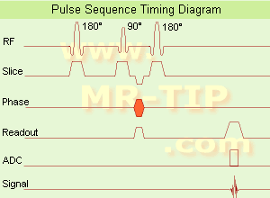
Inversion recovery doubles the distance spins will recover, allowing more time for T1 differences. A 180° preparation pulse inverts the net magnetization to the negative longitudinal magnetization prior to the 90° excitation pulse. This specialized application of the inversion recovery sequence set the inversion time (t) of the sequence at 0.69 times the T1 of fat. The T1 of fat at 1.5 Tesla is approximately 250 with a null point of 170 ms while at 0.5 Tesla its 215 with a 148 ms null point. At the moment of excitation, about 120 to 170 ms after the 180° inversion pulse (depending of the magnetic field) the magnetization of the fat signal has just risen to zero from its original, negative, value and no fat signal is available to be flipped into the transverse plane.
When deciding on the optimal T1 time, factors to be considered include not only the main field strength, but also the tissue to be suppressed and the anatomy. In comparison to a conventional spin echo where tissues with a short T1 are bright due to faster recovery, fat signal is reversed or darkened. Because body fluids have both a long T1 and a long T2, it is evident that STIR offers the possibility of extremely sensitive detection of body fluid. This is of course, only true for stationary fluid such as edema, as the MRI signal of flowing fluids is governed by other factors.

See also Fat Suppression and Inversion Recovery Sequence.

• View the DATABASE results for 'Short T1 Inversion Recovery' (3).Open this link in a new window

 
Further Reading:
  Basics:
Can Short Tau Inversion Recovery (STIR) Imaging Be Used as a Stand-Alone Sequence To Assess a Perianal Fistulous Tract on MRI? A Retrospective Cohort Study Comparing STIR and T1-Post Contrast Imaging
Wednesday, 17 January 2024   by www.cureus.com    
  News & More:
Generating Virtual Short Tau Inversion Recovery (STIR) Images from T1- and T2-Weighted Images Using a Conditional Generative Adversarial Network in Spine Imaging
Wednesday, 25 August 2021
Short tau inversion recovery (STIR) after intravenous contrast agent administration obscures bone marrow edema-like signal on forefoot MRI
Tuesday, 13 July 2021   by www.springermedizin.de    
Turbo Inversion Recovery 
( TIR / TIRM / IR-TSE - Inversion Recovery Turbo Spin Echo / FIR - Fast Inversion Recovery)
A turbo / fast spin echo sequence with long TI for fluid suppression (FLAIR) or with short TI for fat suppression (STIR). This sequence allows for a true inversion recovery display that shows the arithmetic sign of the signal.
TIRM means a turboIR with a magnitude display.

See also Inversion Recovery, Inversion Recovery Sequence and Fast Spin Echo.
 
Further Reading:
  News & More:
Accurate T1 Quantification Using a Breath-hold Inversion Recovery TrueFISP Sequence
2003   by rsna2003.rsna.org    
  Gradient Echo Sequence top
In the beginning the Universe was created. This has made a lot of people very angry and been widely regarded as a bad move.
- Douglas Adams
 
Share This Page
FacebookTwitterLinkedIn

MR-TIP    
Community   
User
Pass
Forgot your UserID/Password ?    


Look
      Ups


MR-TIP.com uses cookies! By browsing MR-TIP.com, you agree to our use of cookies.

Magnetic Resonance - Technology Information Portal
Member of SoftWays' Medical Imaging Group - MR-TIP • Radiology-TIP • Medical-Ultrasound-Imaging • 
Copyright © 2003 - 2024 SoftWays. All rights reserved. [ 23 April 2024]
Terms of Use | Privacy Policy | Advertising
 [last update: 2024-02-26 03:41:00]