 | Info
Sheets |
| | | | | | | | | | | | | | | | | | | | | | | | |
 | Out-
side |
| | | | |
|
| | | | |
Result : Searchterm 'sampling frequency' found in 0 term [ ] and 3 definitions [ ] and 3 definitions [ ], (+ 15 Boolean[ ], (+ 15 Boolean[ ] results ] results
| | 1 - 5 (of 18) nextResult Pages : [1] [2 3 4] |  | |  | Searchterm 'sampling frequency' was also found in the following service: | | | | |
| |  |
| |
|
If the receiving RF coil is sensitive to tissue signal arising from outside the desired FOV, this undesired signal may be incorrectly mapped to a location within the image, a phenomenon known as aliasing. This is a consequence of the acquired k-space frequencies not being sampled densely enough, whereby portions of the object outside of the desired FOV get mapped to an incorrect location inside the FOV.
The sampling frequency should be at least twice the frequency being sampled. The maximum measurable frequency is therefore equal to half the sampling frequency. This is the so-called Nyquist limit. When the frequency is higher than the Nyquist limit, aliasing occurs.
A similar problem occurs in the phase encoding direction, where the phases of signal-bearing tissues outside of the FOV in the y-direction are a replication of the phases that are encoded within the FOV. This signal will be mapped, or wrapped back into the image at incorrect locations, and is seen as artifact.
See also Aliasing Artifact. | | | | | | • Share the entry 'Aliasing':    | | | | | | | | | |  Further Reading: Further Reading: | News & More:
|
|
| |
| | | | | |
| |
|
Oversampling is the increase in data to avoid aliasing and wrap around artifacts. Aliasing is the incorrectly mapping of tissue signal from outside the FOV to a location inside the FOV. This is caused by the fact, that the acquired k-space frequency data is not sampled density enough.
Oversampling in frequency direction, done by increasing the sampling frequency, prevents this aliasing artifact. The proper frequency based on the sampling theorem (Shannon sampling theorem/Nyquist sampling theorem) must be at least twice the frequency of each frequency component in the incoming signal. All frequency components above this limit will be aliased to frequencies between zero and half of the sampling frequency and combined with the proper signal information, which creates the artifact.
Oversampling creates a larger field of view, more data needs to be stored and processed, but this is for modern MRI systems not a real problem. Oversampling in phase direction ( no phase wrap), to eliminate wrap around artifacts, by increasing the number of phase encoding steps, results in longer scan/processing times. | | | |
• View the DATABASE results for 'Oversampling' (10).
| | | | | | Further Reading: | Basics:
|
|
| |
| | | | | |
| |
|
| | | |
• View the DATABASE results for 'Nyquist Limit' (3).
| | | | |
| | | Searchterm 'sampling frequency' was also found in the following service: | | | | |
| |  |
| |
|

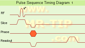
(EPI) Echo planar imaging is one of the early magnetic resonance imaging sequences (also known as Intascan), used in applications like diffusion, perfusion, and functional magnetic resonance imaging. Other sequences acquire one k-space line at each phase encoding step. When the echo planar imaging acquisition strategy is used, the complete image is formed from a single data sample (all k-space lines are measured in one repetition time) of a gradient echo or spin echo sequence (see single shot technique) with an acquisition time of about 20 to 100 ms.
The pulse sequence timing diagram illustrates an echo planar imaging sequence from spin echo type with eight echo train pulses. (See also Pulse Sequence Timing Diagram, for a description of the components.)
In case of a gradient echo based EPI sequence the initial part is very similar to a standard gradient echo sequence. By periodically fast reversing the readout or frequency encoding gradient, a train of echoes is generated.
EPI requires higher performance from the MRI scanner like much larger gradient amplitudes. The scan time is dependent on the spatial resolution required, the strength of the applied gradient fields and the time the machine needs to ramp the gradients.
In EPI, there is water fat shift in the phase encoding direction due to phase accumulations. To minimize water fat shift (WFS) in the phase direction fat suppression and a wide bandwidth (BW) are selected. On a typical EPI sequence, there is virtually no time at all for the flat top of the gradient waveform. The problem is solved by "ramp sampling" through most of the rise and fall time to improve image resolution.
The benefits of the fast imaging time are not without cost. EPI is relatively demanding on the scanner hardware, in particular on gradient strengths, gradient switching times, and receiver bandwidth. In addition, EPI is extremely sensitive to image artifacts and distortions. | | | |
• View the DATABASE results for 'Echo Planar Imaging' (19).
| | |
• View the NEWS results for 'Echo Planar Imaging' (1).
| | | | | | Further Reading: | Basics:
|
|
| |
| | | | | |
| |
|
| | | |
• View the DATABASE results for 'Pulse Sequence Timing Diagram' (7).
| | | | |
| | | | |
| | |
| | | 1 - 5 (of 18) nextResult Pages : [1] [2 3 4] |
| |
|
| |
 | Look
Ups |
| |


 The schematic figures of a
 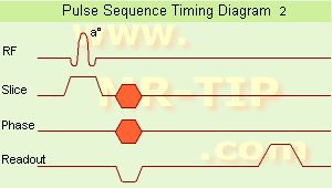 The
The 