 | Info
Sheets |
| | | | | | | | | | | | | | | | | | | | | | | | |
 | Out-
side |
| | | | |
|
| | | | |
Result : Searchterm 'high field' found in 1 term [ ] and 35 definitions [ ] and 35 definitions [ ] ]
| | 1 - 5 (of 36) nextResult Pages : [1] [2 3 4 5 6 7 8] |  | |  | Searchterm 'high field' was also found in the following services: | | | | |
| |  |
| |
|
The principal advantage of MRI at high field is the increase in signal to noise ratio. This can be used to improve anatomic and/or temporal resolution and reduce scan time while preserving image quality. MRI devices for whole body imaging for human use are available up to 3 tesla (3T). Functional MRI ( fMRI) and MR spectroscopy ( MRS) benefit significantly. In addition, 3T machines have a great utility in applications such as TOF MRA and DTI. Higher field strengths are used for imaging of small parts of the body or scientific animal experiments. Higher contrast may permit reduction of gadolinium doses and, in some cases, earlier detection of disease.
Using high field MRI//MRS, the RF-wavelength and the dimension of the human body complicating the development of MR coils. The absorption of RF power causes heating of the tissue. The energy deposited in the patient's tissues is fourfold higher at 3T than at 1.5T. The specific absorption rate (SAR) induced temperature changes of the human body are the most important safety issue of high field MRI//MRS.
Susceptibility and chemical shift dispersion increase like T1, therefore high field MRI occasionally exhibits imaging artifacts. Most are obvious and easily recognized but some are subtle and mimic diseases. A thorough understanding of these artifacts is important to avoid potential pitfalls. Some imaging techniques or procedures can be utilized to remove or identify artifacts. See also Diffusion Tensor Imaging.
See also the related poll result: ' In 2010 your scanner will probably work with a field strength of' | | | | | | | | • Share the entry 'High Field MRI':    | | | | | | | | | |  Further Reading: Further Reading: | | Basics:
|
|
News & More:
| |
| |
| | | | |  |
| |
|

From ISOL Technology
'Ultra high field MR system, it's right close to you.
FORTE 3.0T is the new standard for the future ultra high field MR system.
If you are pushing the limits of your existing clinical MR scanner, the FORTE will surely take you to the next level of diagnostic imaging.
FORTE is the core leader of the medical technology in the 21st century. Proving effects of fMRI that cannot be measured with MRI less than 2.0T.'
Device Information and Specification
CLINICAL APPLICATION
Whole body
CONFIGURATION
Short bore compact
128 x 128, 256 x 256, 512 x 512, 1024 x 1024
| | | |
• View the DATABASE results for 'FORTE 3.0T™' (2).
| | | | |
| | | | | |
| |
|
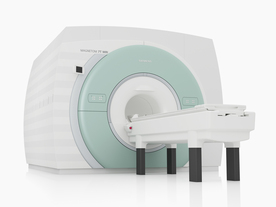
From Siemens Medical Systems;
The MAGNETOM 7T is designed as an open research platform. 7T MRI provides anatomical detail at the submillimiter scale, enhanced contrast mechanisms, outstanding spectroscopy performance, ultra-high resolution functional imaging ( fMRI), multinuclear whole-body MRI and functional information.
This ultra high field (UHF) MRI device is a research system and not cleared, approved or licensed in any jurisdiction for patient examinations.
Device Information and Specification
CLINICAL APPLICATION
Whole body
High-performance, ultra high field coils available. Integration and support for coil developments.
CHANNELS (min. / max. configuration)
32, optional 8 channels TX array
40 x 40 x 30 cm³ less than 8% nonlinearity
MAGNET WEIGHT (gantry included)
35017 kg
DIMENSION H*W*D (gantry included)
320 x 240 x 317,5 cm
MAX. AMPLITUDE
up to 70 mT/m
Up to 3rd order shim coils, user configurable B0 shim ? B0 maps and ROI definition
POWER REQUIREMENTS
2000 Volts, 650A
| | | | | | | Further Reading: | Basics:
|
|
News & More:
| |
| |
| | | Searchterm 'high field' was also found in the following services: | | | | |
| | |
| |
|

From ONI Medical Systems, Inc.;
MSK-Extreme™ MRI system is a dedicated high field extremity imaging device, designed to provide orthopedic surgeons and other physicians with detailed diagnostic images of the foot, ankle, knee, hand, wrist and elbow, all with the clinical confidence and advantages derived from high field, whole body MRI units. The light weight (less than 650 kg) of the OrthOne System performs rapid patient studies, is easy to operate, has a patient friendly open environment and can be installed in a practice office or hospital, all at a cost similar to a low field extremity machine.
New features include a more powerful operating system that offers increased scan speed as well as a 160-mm knee coil with higher signal to noise ratio, and the option of a CD burner.
Device Information and Specification 16 cm knee, 18 cm lower extremity;; 12.3 cm upper extremity, additional high resolution v-SPEC Coils: 80 mm, 100 mm, or 145 mm. SE, FSE, GE2D, GE3D, Inversion recovery (IR), Driven Equilibrium, Fat Saturation (FS), STIR, MT, PD, Flow Compensation (FC), RF spoiling, MTE, No Phase Wrap (NPW) IMAGING MODES Scout, single, multislice, volume 2D less than 200 msec/image X/Y: 64-512; 2 pixel steps 4,096 grey lvls; 256 lvls in 3D POWER REQUIREMENTS 115VAC, 1phase, 20A; 208VAC, 3 phase, 30A COOLING SYSTEM TYPE LHe with 2 stage cold head 1.25m radial x 1.8m axial | | | | | | | Further Reading: | Basics:
|
|
| |
| | | | | |
| |
|
Open MRI scanners have been developed for people who are anxious or obese or for examination of small parts of the body, such as the extremities ( knee, shoulder). In addition, some systems offer imaging in different positions and sequences of movements.
The basic technology of an open MRI machine is similar to that of a traditional MRI device.
The major difference for the patient is that instead of lying in a narrow tunnel, the imaging table has more space around the body so that the magnet does not completely surround the person being tested.
Types of constructions:
•
Semi open high field MRI scanners provide an ultra short bore (tunnel) and widely flared ends. In this type of MRI systems, patients lie with the head in the space outside the bore, if for example the hips are examined.
•
Open low field MRI machines have often a wide open design, e.g. an open C-arm scanner is shaped like two large discs separated by a large pillar. Patients have an open sided feeling and more space around them allows a wider range of positions.
•
Advanced open MRI scanners combine the advantages of both, the high field strength, newest gradient technology and wide open design. Even scans of patients in upright, weight-bearing positions are possible (e.g. Upright™ MRI formerly Stand-Up MRI).
Difficulties with a traditional MRI scan include claustrophobia and patient size or, for health related reasons, patients who are not able to receive this type of diagnostic test. The MRI unit is a limited space, and some patients may be too large to fit in a narrow tunnel. In addition, weight limits can restrict the use of some scanners. The open MRI magnet has become the best option for those patients.
All of the highest resolution MRI scanners are tunnels and tend to accentuate the claustrophobic reaction. While patients may find the open MRI scanners easier to tolerate, some machines use a lower field magnet and generates lower image quality or have longer scan time. The better performance of an advanced open MRI scanner allows good image quality caused by the higher signal to noise ratio with maximum patient comfort.
See also Claustrophobia, MRI scan and Knee MRI. | | | |
• View the DATABASE results for 'Open MRI' (37).
| | |
• View the NEWS results for 'Open MRI' (16).
| | | | | | Further Reading: | Basics:
|
|
News & More:
| |
| |
| | | | |
| | | |
|
| |
 | Look
Ups |
| |


