Magnetic Resonance - Technology Information Portal Welcome to MRI Technology
Info
  Sheets

Out-
      side


 'cardiac' 
SEARCH FOR    
 
  2 3 5 A B C D E F G H I J K L M N O P Q R S T U V W X Y Z
Result : Searchterm 'cardiac' found in 11 terms [] and 75 definitions []
previous     51 - 55 (of 86)     next
Result Pages : [1 2 3]  [4 5 6 7 8 9 10 11 12 13 14 15 16 17 18]
Searchterm 'cardiac' was also found in the following services: 
spacer
News  (82)  Resources  (27)  Forum  (13)  
 
Gating
 
Synchronization of imaging with a phase of the cardiac or respiratory cycles.
A variety of means for detecting these cycles can be used, such as the ECG, peripheral pulse, chest motion, etc. The synchronization can be prospective or retrospective.
spacer
 
• Related Searches:
    • Trigger Delay
    • Cardiac MRI
    • Fast Relaxation Fast Spin Echo
    • Cine Sequence
    • Pulse Triggering
 
Further Reading:
  Basics:
Electrocardiogram in an MRI Environment: Clinical Needs, Practical Considerations, Safety Implications, Technical Solutions and Future Directions
Wednesday, 25 January 2012   by cdn.intechopen.com    
MRI Resources 
MRI Technician and Technologist Career - Lung Imaging - Sequences - Universities - Spectroscopy pool - Used and Refurbished MRI Equipment
 
Imaging CoilInfoSheet: - Coils - 
Intro, 
Overview, 
etc.MRI Resource Directory:
 - Coils -
 
Imaging coils are radio frequency coils used in magnetic resonance imaging for sending and/or receiving electromagnetic radiation. Several MR imaging coils in different shapes and sizes are necessary for different body parts and to handle individual applications.
For Example:
Birdcage coils provide high homogeneity and good signal to noise ratio (SNR) in brain MRI scans.
Surface coils with small coil diameter and higher SNR enable to image the temporomandibular joints (TMJ).
The implemented body coil allows the scanning of body parts with large field of views.
Micro coils are available to image finger joints.
High performance phased array coils are today state-of-the-art for a wide range of applications from head, spine, knee or shoulder to cardiac MRI.
For different types of coils see Volume Coil, Sense Coil, Array Coil, Surface Coil, and Bird Cage Coil.

See also the related poll result: '3rd party coils are better than the original manufacturer coils'
spacer

• View the DATABASE results for 'Imaging Coil' (7).Open this link in a new window


• View the NEWS results for 'Imaging Coil' (9).Open this link in a new window.
 
Further Reading:
  Basics:
Radio-frequency Coil Selection for MR Imaging of the Brain and Skull Base1
   by radiology.rsnajnls.org    
MRI Resources 
Supplies - - Liver Imaging - Breast MRI - MRCP - Artifacts
 
ImplantsForum -
related threadsMRI Resource Directory:
 - Safety -
 
Implants that involve magnets such as magnetic sphincters, stoma plugs, dental implants, etc., can be demagnetized by the MRI device. They should be removed prior to the examination.
mri safety guidance
MRI Safety Guidance
A particular danger is presented by small metallic surgical implants. Haemostatic or other clips in the CNS can move in their position. Dislocation by magnetic attraction or torque presents a risk in MRI examinations. There is a minimal risk in other parts of the body, because after the healing phase of six to eight weeks, fibrosis and encasement of the clip help to keep it in a stable position.
The label stainless steel is not a guarantee for non-ferromagnetic steel.

See also Cardiac Pacemaker and MRI Safety.
spacer

• View the DATABASE results for 'Implants' (13).Open this link in a new window


• View the NEWS results for 'Implants' (2).Open this link in a new window.
 
Further Reading:
  Basics:
Newer Heart Devices Safe During MRI
Monday, 23 August 2004   by www.hospimedica.com    
FDA Releases New Guidance On Establishing Safety, Compatibility Of Passive Implants In MR Environments
Tuesday, 16 December 2014   by www.meddeviceonline.com    
Pain, magnet displacement in MRI in patients with cochlear implants
Thursday, 20 November 2014   by medicalxpress.com    
  News & More:
On the estimation of the worst-case implant-induced RF-heating in multi-channel MRI.
Thursday, 2 March 2017   by www.ncbi.nlm.nih.gov    
Positive diagnosis for neural therapeutic implants
Tuesday, 19 April 2016   by medicalxpress.com    
ITOCHU Named the Exclusive Distributor for ViewRay's MRI-Guided Radiation Therapy System in Japan
Thursday, 22 January 2015   by www.prnewswire.com    
FDA grants PMA-Supplement approval for Flowonix Medical's Prometra II system
Tuesday, 20 January 2015   by www.medicaldevice-network.com    
FDA Approves MED-EL's SYNCHRONY Cochlear Implant
Friday, 23 January 2015   by www.businesswire.com    
Searchterm 'cardiac' was also found in the following services: 
spacer
News  (82)  Resources  (27)  Forum  (13)  
 
Infinion 1.5TPanorama 0.2InfoSheet: - Devices -
Intro, 
Types of Magnets, 
Overview, 
etc.MRI Resource Directory:
 - Devices -
 
From Philips Medical Systems; www.medical.philips.com/main/products/mri/products/infinion1.5t/features/ 
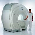
Philips Infinion 1.5 T is designed to maximize the efficiency and quality of patient care. Developed with the patient in mind, the Infinion is the shortest and most open 1.5T scanner available. The unique 'ultra short' 1.4 m magnet assures patient comfort and acceptance without compromising image quality and clinical performance.
Device Information and Specification
CLINICAL APPLICATION
Whole body
CONFIGURATION
Ultra short bore
Head, head / neck, integrated C-spine, L/T spine array, small large GP coils, body flex array, torso pelvis array, breast array, endocavitary, shoulder array, lower extremity, hand / wrist, cardiac, PV array
SYNCHRONIZATION
ECG/peripheral, respiratory gating
PULSE SEQUENCES
SE, TSE, SS TSE, EPI, IR, STIR, FLAIR, FFE, TFE, T1 TFE, T2 TFE, Presat, Fatsat, MTC, Diff-opt., Angiography: PCA, MCA, TOF
IMAGING MODES
Single slice, single volume, multi slice, multi volume
TR
3.1 msec minimum
TE
0.9 msec minimum
SINGLE/MULTI SLICE
80 images/sec std.; up to320 opt.@256
FOV
0.4 - 56 cm
2D: 0.3 mm; 3D: 0.2 mm
1024 x 1024
MEASURING MATRIX
8 x 8 to 1024 x 1024
PIXEL INTENSITY
256 gray scale
BORE DIAMETER
or W x H
60 cm diameter (patient)
MAGNET WEIGHT
4100 kg w/cryogen's
H*W*D
233 (lead fitted) x 198 x 140 cm
POWER REQUIREMENTS
400/480 V
COOLING SYSTEM TYPE
Closed loop, chilled water
CRYOGEN USE
0.06 L/hr helium
STRENGTH
30 mT/m
5-GAUSS FRINGE FIELD
3.0 m / 5.0 m
Passive/active
spacer

• View the DATABASE results for 'Infinion 1.5T™' (2).Open this link in a new window

MRI Resources 
Contrast Enhanced MRI - Chemistry - IR - Service and Support - Portals - Journals
 
Intera 0.5TPanorama 0.2InfoSheet: - Devices -
Intro, 
Types of Magnets, 
Overview, 
etc.MRI Resource Directory:
 - Devices -
 
www.medical.philips.com/de/products/mri/products/ From Philips Medical Systems;
this low field MRI system of the Intera-family is onto diagnostic safety and flexibility aligned - cost effective and with high patient acceptance.
Device Information and Specification
CLINICAL APPLICATION
Whole body
CONFIGURATION
Short bore compact
Standard: head, body, C1, C3; Optional: Small joint, flex-E, flex-R, endocavitary (L and S), dual TMJ, knee, neck, T/L spine, breast; Optional phased array: Spine, pediatric, 3rd party connector, Flex-S-M-L, flex body, flex cardiac, neuro-vascular, head
SYNCHRONIZATION
ECG/peripheral: Optional/yes, respiratory gating
PULSE SEQUENCES
SE, Modified-SE (TSE), DAVE, STIR, FLAIR, SPIR, MTC, Dynamic, Keyhole, CLEAR, Q Flow, Balanced FFE, Multi Chunk 3D, Multi Stack 3D, FFE-EPI, SE-EPI, IR-EPI, GRASE, Diffusion Imaging, Perfusion Imaging;; Angiography: Inflow MRA, TONE, PCA, CE MRA
IMAGING MODES
Single Slice 2D , Multi Single Slice 2D, Multi Slice 2D, 3D, Multi Chunk 3D, Multi Stack 3D
TR
Min. 2.9 (Omni) msec
TE
Min. 1.0 (Omni) msec
SINGLE/MULTI SLICE
RapidView Recon. greater than 500 @ 256 Matrix
FOV
Max. 53 cm
0.1 mm (Omni)
128 x 128, 256 x 256,512 x 512,1024 x 1024
MEASURING MATRIX
Variable in 1% increments
PIXEL INTENSITY
Lum.: 120 cd/m2; contrast: 150:1
Variable (op. param. depend.)
60 x 60 cm
MAGNET WEIGHT
2500 kg
H*W*D
240 x 188 x 157 cm
POWER REQUIREMENTS
380/400 V
CRYOGEN USE
0.03 L/hr helium
STRENGTH
23 mT/m (Omni)
5-GAUSS FRINGE FIELD
2.1 m / 2.8 m
Passive and dynamic
spacer

• View the DATABASE results for 'Intera 0.5T™' (2).Open this link in a new window

MRI Resources 
MRCP - Pediatric and Fetal MRI - Research Labs - MRI Training Courses - Directories - Spectroscopy
 
previous      51 - 55 (of 86)     next
Result Pages : [1 2 3]  [4 5 6 7 8 9 10 11 12 13 14 15 16 17 18]
 Random Page
 
Share This Page
FacebookTwitterLinkedIn

MR-TIP    
Community   
User
Pass
Forgot your UserID/Password ?    


Next big thing in MRI will be :
AI 
remote operator 
personalized protocols 
helium-free 
molecular MRI 
portable MRI 

Look
      Ups


MR-TIP.com uses cookies! By browsing MR-TIP.com, you agree to our use of cookies.

Magnetic Resonance - Technology Information Portal
Member of SoftWays' Medical Imaging Group - MR-TIP • Radiology-TIP • Medical-Ultrasound-Imaging • 
Copyright © 2003 - 2024 SoftWays. All rights reserved. [ 25 April 2024]
Terms of Use | Privacy Policy | Advertising
 [last update: 2024-02-26 03:41:00]