Magnetic Resonance - Technology Information Portal Welcome to MRI Technology
Info
  Sheets

Out-
      side


 'Transverse Relaxation Time' 
SEARCH FOR    
 
  2 3 5 A B C D E F G H I J K L M N O P Q R S T U V W X Y Z
Result : Searchterm 'Transverse Relaxation Time' found in 1 term [] and 3 definitions [], (+ 12 Boolean[] results
1 - 5 (of 16)     next
Result Pages : [1]  [2 3 4]
Searchterm 'Transverse Relaxation Time' was also found in the following service: 
spacer
News  (1)  
 
Transverse Relaxation Time
 
The T2 time constant, which determines the rate at which excited protons reach equilibrium, or go out of phase with each other. A measure of the time taken for spinning protons to lose phase coherence among the nuclei spinning perpendicular to the main field due to interaction between spins, resulting in a reduction in the transverse magnetization. The transverse magnetization value will drop from maximum to a value of about 37% of its original value in a time of T2.
 
Images, Movies, Sliders:
 Brain MRI Images Axial T2  Open this link in a new window
      

 Breast MRI 2 Transverse T2 001  Open this link in a new window
 
spacer
 
• Share the entry 'Transverse Relaxation Time':  Facebook  Twitter  LinkedIn  
MRI Resources 
Case Studies - Safety Training - Safety pool - Shielding - Breast Implant - Non-English
 
Lung ImagingMRI Resource Directory:
 - Lung Imaging -
 
Lung imaging is furthermore a challenge in MRI because of the predominance of air within the lungs and associated susceptibility issues as well as low signal to noise of the inflated lung parenchyma. Cardiac and respiratory triggered or breath hold sequences allow diagnostic imaging, however a comparable image quality with computed tomography is still difficult to achieve.
Assumptions for lung MRI:
Low signal to noise ratio of the inherently low lung proton density.
Cardiac and respiratory motion artifacts.
Magnetic susceptibility effects of large magnetic field gradients.
Very short transverse relaxation times and significant diffusion yielding short T2 (30-70 msec), short T2* (1-3 msec), and additional long T1 relaxation times (1300-1500 msec).
The extreme short T2 values are responsible for a fast signal decay during a single shot readout, resulting in blurring.

The current trends in MRI are the use of new imaging technologies and increasingly powerful magnetic fields. Among these technologies are parallel imaging techniques as well as ventilation agents like hyperpolarized helium for the use as an inert inhalational contrast agent to study lung ventilation properties. With hyperpolarized gases clear images of the lungs can be obtained without using a large magnetic field (see also back projection imaging). Single shot sequences (e.g. TSE or Half Fourier Acquisition Single Shot Turbo Spin Echo HASTE) used in lung MR imaging benefits from parallel imaging techniques due to reduced relaxation time effects during the echo train and therefore reduced image blurring as well as reduced motion artifacts.
In the future, more effective contrast agents may provide an alternative solution to the need for high field MRI. Dynamic contrast enhanced MRI perfusion has demonstrated a potential in the diagnosis of pulmonary embolism or to characterize lung cancer and mediastinal tumors. 3D contrast enhanced magnetic resonance angiography of the thoracic vessel.

See also the related poll result: 'MRI will have replaced 50% of x-ray exams by'
 
Images, Movies, Sliders:
 Anatomic Imaging of the Lungs  Open this link in a new window
      

Courtesy of  Robert R. Edelman
 Normal Lung Gd Perfusion MRI  Open this link in a new window
      

Courtesy of  Robert R. Edelman

 MRI Thorax Basal Plane  Open this link in a new window
 
Radiology-tip.comradLung Scintigraphy
spacer

• View the DATABASE results for 'Lung Imaging' (7).Open this link in a new window


• View the NEWS results for 'Lung Imaging' (3).Open this link in a new window.
 
Further Reading:
  Basics:
A safer approach for diagnostic medical imaging
Monday, 29 September 2014   by www.eurekalert.org    
Parallel Lung Imaging(.pdf)
  News & More:
Chest MRI a viable alternative to chest CT in COVID-19 pneumonia follow-up
Monday, 21 September 2020   by www.healthimaging.com    
CT Imaging Features of 2019 Novel Corona virus (2019-nCoV)
Tuesday, 4 February 2020   by pubs.rsna.org    
Polarean Imaging Phase III Trial Results Point to Potential Improvements in Lung Imaging
Wednesday, 29 January 2020   by www.diagnosticimaging.com    
Low Power MRI Helps Image Lungs, Brings Costs Down
Thursday, 10 October 2019   by www.medgadget.com    
Chest MRI Using Multivane-XD, a Novel T2-Weighted Free Breathing MR Sequence
Thursday, 11 July 2019   by www.sciencedirect.co    
Researchers Review Importance of Non-Invasive Imaging in Diagnosis and Management of PAH
Wednesday, 11 March 2015   by lungdiseasenews.com    
New MRI Approach Reveals Bronchiectasis' Key Features Within the Lung
Thursday, 13 November 2014   by lungdiseasenews.com    
MRI techniques improve pulmonary embolism detection
Monday, 19 March 2012   by medicalxpress.com    
  News & More:
Partnership with VIDA to streamline adoption of advanced MRI of the lungs
Monday, 11 September 2023   by www.itnonline.com    
MRI Resources 
Societies - Safety Products - Examinations - MRI Physics - Research Labs - DICOM
 
Spin Spin Relaxation Time
 
The spin spin relaxation time is the time the spins need to dephase in the transverse plane.
See T2 Time, and Transverse Relaxation Time.
spacer

• View the DATABASE results for 'Spin Spin Relaxation Time' (2).Open this link in a new window

 
Further Reading:
  News & More:
MULTIEXPONENTIAL PROTON SPIN-SPIN RELAXATION IN MAGNETIC RESONANCE IMAGING OF HUMAN BRAIN TUMORS
Friday, 26 March 1999   by www.dkfz-heidelberg.de    
T2* cardiac MRI allows prediction of severe reperfusion injury after STEMI
Tuesday, 9 November 2010   by www.medwire-news.md    
Searchterm 'Transverse Relaxation Time' was also found in the following service: 
spacer
News  (1)  
 
T2 TimeForum -
related threads
 
The T2 relaxation time (spin spin relaxation time or transverse relaxation time), is a biological parameter that is used in MRIs to distinguish between tissue types and is termed 'Time 2' or T2. It is a tissue-specific time constant for protons and is dependent on the exchanging of energy with near by nuclei. T2 weighted images rely upon local dephasing of spins following the application of the transverse energy pulse. T2 is the decay of magnetization perpendicular to the main magnetic field (in an ideal homogeneous field).
Due to interaction between the spins, they lose their phase coherence, which results in a loss of transverse magnetization and MRI signal. After time T2 transverse magnetization has lost 63% of its original value. This tissue parameter determines the contrast.
The T2 relaxation is temperature dependent. At a lower temperature molecular motion is reduced and the decay times are reduced.
Fat has a very efficient energy exchange and therefore it has a relatively short T2.
Water is less efficient than fat in the exchange of energy, and therefore it has a long T2 time.

See also T2 Weighted Image and Magnetic Resonance Imaging MRI.
 
Images, Movies, Sliders:
 Breast MRI Images T2 And T1  Open this link in a new window
      
 MRI of the Skull Base  Open this link in a new window
    
SlidersSliders Overview

 Knee MRI Transverse 002  Open this link in a new window
    
 
spacer

• View the DATABASE results for 'T2 Time' (16).Open this link in a new window

 
Further Reading:
  News & More:
MYELIN-SELECTIVE MRI: PULSE SEQUENCE DESIGN AND OPTIMIZATION
   by www.imaging.robarts.ca    
MRI Resources 
Jobs pool - Homepages - Process Analysis - Spine MRI - Safety pool - Fluorescence
 
Spin Echo SequenceInfoSheet: - Sequences - 
Intro, 
Overview, 
Types of, 
etc.MRI Resource Directory:
 - Sequences -
 
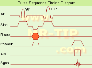
Spin Echo Timing Diagram (SE) The most common pulse sequence used in MR imaging is based of the detection of a spin or Hahn echo. It uses 90° radio frequency pulses to excite the magnetization and one or more 180° pulses to refocus the spins to generate signal echoes named spin echoes (SE).
In the pulse sequence timing diagram, the simplest form of a spin echo sequence is illustrated.
The 90° excitation pulse rotates the longitudinal magnetization (Mz) into the xy-plane and the dephasing of the transverse magnetization (Mxy) starts.
The following application of a 180° refocusing pulse (rotates the magnetization in the x-plane) generates signal echoes. The purpose of the 180° pulse is to rephase the spins, causing them to regain coherence and thereby to recover transverse magnetization, producing a spin echo.
The recovery of the z-magnetization occurs with the T1 relaxation time and typically at a much slower rate than the T2-decay, because in general T1 is greater than T2 for living tissues and is in the range of 100-2000 ms.
The SE pulse sequence was devised in the early days of NMR days by Carr and Purcell and exists now in many forms: the multi echo pulse sequence using single or multislice acquisition, the fast spin echo (FSE/TSE) pulse sequence, echo planar imaging (EPI) pulse sequence and the gradient and spin echo (GRASE) pulse sequence;; all are basically spin echo sequences.
In the simplest form of SE imaging, the pulse sequence has to be repeated as many times as the image has lines.
Contrast values:
PD weighted: Short TE (20 ms) and long TR.
T1 weighted: Short TE (10-20 ms) and short TR (300-600 ms)
T2 weighted: Long TE (greater than 60 ms) and long TR (greater than 1600 ms)
With spin echo imaging no T2* occurs, caused by the 180° refocusing pulse. For this reason, spin echo sequences are more robust against e.g., susceptibility artifacts than gradient echo sequences.

See also Pulse Sequence Timing Diagram to find a description of the components.
 
Images, Movies, Sliders:
 Shoulder Coronal T1 SE  Open this link in a new window
    

Courtesy of  Robert R. Edelman
 Shoulder Axial T1 SE  Open this link in a new window
 MRI Orbita T1  Open this link in a new window
    
 
spacer

• View the DATABASE results for 'Spin Echo Sequence' (24).Open this link in a new window

 
Further Reading:
  Basics:
Fast Spin Echo(.pdf)
Tuesday, 24 January 2006   by www.81bones.net    
Magnetic resonance imaging
   by www.scholarpedia.org    
FUNDAMENTALS OF MRI: Part I
   by www.e-radiography.net    
  News & More:
New MR sequence helps radiologists more accurately evaluate abnormalities of the uterus and ovaries
Thursday, 23 April 2009   by www.eurekalert.org    
MRI techniques improve pulmonary embolism detection
Monday, 19 March 2012   by medicalxpress.com    
MRI Resources 
Image Quality - Musculoskeletal and Joint MRI - NMR - Stent - Software - Spectroscopy pool
 
     1 - 5 (of 16)     next
Result Pages : [1]  [2 3 4]
 Random Page
 
Share This Page
FacebookTwitterLinkedIn

MR-TIP    
Community   
User
Pass
Forgot your UserID/Password ?    


Next big thing in MRI will be :
AI 
remote operator 
personalized protocols 
helium-free 
molecular MRI 
portable MRI 

Look
      Ups


MR-TIP.com uses cookies! By browsing MR-TIP.com, you agree to our use of cookies.

Magnetic Resonance - Technology Information Portal
Member of SoftWays' Medical Imaging Group - MR-TIP • Radiology-TIP • Medical-Ultrasound-Imaging • 
Copyright © 2003 - 2024 SoftWays. All rights reserved. [ 16 April 2024]
Terms of Use | Privacy Policy | Advertising
 [last update: 2024-02-26 03:41:00]