Magnetic Resonance - Technology Information Portal Welcome to MRI Technology
Info
  Sheets

Out-
      side


 'Tesla' 
SEARCH FOR    
 
  2 3 5 A B C D E F G H I J K L M N O P Q R S T U V W X Y Z
Result : Searchterm 'Tesla' found in 3 terms [] and 38 definitions []
previous     21 - 25 (of 41)     next
Result Pages : [1]  [2 3 4 5 6 7 8 9]
Searchterm 'Tesla' was also found in the following services: 
spacer
News  (56)  Resources  (12)  Forum  (14)  
 
High Field MRI
 
The principal advantage of MRI at high field is the increase in signal to noise ratio. This can be used to improve anatomic and/or temporal resolution and reduce scan time while preserving image quality. MRI devices for whole body imaging for human use are available up to 3 tesla (3T). Functional MRI (fMRI) and MR spectroscopy (MRS) benefit significantly. In addition, 3T machines have a great utility in applications such as TOF MRA and DTI. Higher field strengths are used for imaging of small parts of the body or scientific animal experiments. Higher contrast may permit reduction of gadolinium doses and, in some cases, earlier detection of disease.
Using high field MRI//MRS, the RF-wavelength and the dimension of the human body complicating the development of MR coils. The absorption of RF power causes heating of the tissue. The energy deposited in the patient's tissues is fourfold higher at 3T than at 1.5T. The specific absorption rate (SAR) induced temperature changes of the human body are the most important safety issue of high field MRI//MRS.
Susceptibility and chemical shift dispersion increase like T1, therefore high field MRI occasionally exhibits imaging artifacts. Most are obvious and easily recognized but some are subtle and mimic diseases. A thorough understanding of these artifacts is important to avoid potential pitfalls. Some imaging techniques or procedures can be utilized to remove or identify artifacts.

See also Diffusion Tensor Imaging.

See also the related poll result: 'In 2010 your scanner will probably work with a field strength of'
Medical-Ultrasound-Imaging.comMagnetic Resonance Guided Focused Ultrasound,  High Intensity Focused Ultrasound
spacer
 
• Related Searches:
    • Contrast to Noise Ratio
    • B0
    • Field Strength
    • Device
    • Signal Intensity
 
Further Reading:
  Basics:
Next-generation 7 T scanner ramps the resolution of brain MR imaging
Wednesday, 17 January 2024   by physicsworld.com    
A paired dataset of T1- and T2-weighted MRI at 3 Tesla and 7 Tesla
Thursday, 27 July 2023   by www.nature.com    
CLINICAL WHOLE BODY MRI AT 3.0 T(.pdf)
2001
Musculoskeletal MRI at 3.0 T: Relaxation Times and Image Contrast
Sunday, 1 August 2004   by www.ajronline.org    
  News & More:
How safe is 7T MRI for patients with neurosurgical implants?
Thursday, 17 November 2022   by healthimaging.com    
Impact of Magnetic Field Inhomogeneity on the Quality of Magnetic Resonance Images and Compensation Techniques: A Review
Saturday, 1 October 2022   by www.dovepress.com    
7-T clinical MRI of the shoulder in patients with suspected lesions of the rotator cuff
Friday, 7 February 2020   by eurradiolexp.springeropen.com    
A 100-hour MRI scan captured the most detailed look yet at a whole human brain
Monday, 8 July 2019   by www.sciencenews.or    
T2-Weighted Liver MRI Using the MultiVane Technique at 3T: Comparison with Conventional T2-Weighted MRI
Friday, 16 October 2015   by www.ncbi.nlm.nih.gov    
Ultra-high-field MRI reveals language centres in the brain in much more detail
Tuesday, 28 October 2014   by medicalxpress.com    
Turbo-FLASH Based Arterial Spin Labeled Perfusion MRI at 7 T
Thursday, 20 June 2013   by www.plosone.org    
High-Resolution, Spin-Echo BOLD, and CBF fMRI at 4 and 7 T(.pdf)
October 2002   by otg.downstate.edu    
Vascular Filters of Functional MRI: Spatial Localization Using BOLD and CBV Contrast
Searchterm 'Tesla' was also found in the following services: 
spacer
Radiology  (4) Open this link in a new windowUltrasound  (1) Open this link in a new window
IMRISâ„¢MRI Resource Directory:
 - Manufacturers -
 
IMRIS® is a Canadian manufacturer of Magnetic Resonance Imaging Systems for use in neurosurgical operating rooms. The 1.5T intra-operative system, developed by IMRIS, is the only system of its kind in the world. This patented, intra-operative system, is designed so that the magnet moves over the patient for imaging (before, during and after surgery), and then is retracted to allow complete surgical access to the patient.

MRI Scanners:

Contact Information
MAIL
IMRIS®
435 Ellice Avenue
Winnipeg.MB
Canada R3B1Y6
PHONE
1-88-304-0114 or 204-983-6867
FAX
204-984-1850
ONLINE
spacer
MRI Resources 
Manufacturers - Open Directory Project - General - Musculoskeletal and Joint MRI - Spectroscopy pool - Functional MRI
 
Intera Achieva 3.0Tâ„¢InfoSheet: - Devices -
Intro, 
Types of Magnets, 
Overview, 
etc.MRI Resource Directory:
 - Devices -
 
www.medical.philips.com/main/products/mri/products/intera_achieva3.0t/ From Philips Medical Systems;
Philips continues to expand the frontiers of utra high field MRI with the introduction of the new Intera Achieva 3.0T™. Its powerful future-safe platform shares all the advantages of the Achieva family and covers applications throughout the whole body.
Device Information and Specification
CLINICAL APPLICATION
Whole body
CONFIGURATION
Short bore compact
Standard: T/R-head, Body, Optional phased array: CTL spine; Optional SENSE coils: Torso, Cardiac, Head, Knee, Breast, Neurovascular, Flex M, Flex L
Optional
SYNCHRONIZATION
ECG/peripheral: Optional/yes, respiratory gating
PULSE SEQUENCES
SE, Modified-SE, IR (T1, T2, PD), STIR, FLAIR, SPIR, FFE, T1-FFE, T2-FFE, Balanced FFE, TFE, Balanced TFE, Dynamic, Keyhole, 3D, Multi Chunk 3D, Multi Stack 3D, K Space Shutter, MTC, TSE, Dual IR, DRIVE, EPI, Cine, 2DMSS, DAVE, Mixed Mode; Angiography: Inflow MRA, TONE, PCA, CE MRA
IMAGING MODES
Single Slice 2D , Multi Single Slice 2D, Multi Slice 2D, 3D, Multi Chunk 3D, Multi Stack 3D
Minimum TR
1.1 msec
FOV
Over 40 cm
0.05 mm
128 x 128, 256 x 256,512 x 512,1024 x 1024 (64 for Bold img)
MEASURING MATRIX
Variable in 1% increments
PIXEL INTENSITY
Lum.: 120 cd/m2; contrast: 150:1
Variable (op. param. depend.)
35 x 35 cm
MAGNET WEIGHT
5500 kg
H*W*D
206 x 200 x 167 cm
POWER REQUIREMENTS
380/400 V
CRYOGEN USE
0.15 L/hr helium
STRENGTH
up to 80 mT/m
5-GAUSS FRINGE FIELD
3.0 m / 5.2 m
Passive and dynamic, 1st order std./2nd opt.
spacer

• View the DATABASE results for 'Intera Achieva 3.0T™' (2).Open this link in a new window

 
Further Reading:
  News & More:
Faster speed, better spatial resolution lead 3T benefits
   by sipi.usc.edu    
Searchterm 'Tesla' was also found in the following services: 
spacer
News  (56)  Resources  (12)  Forum  (14)  
 
Larmor Frequency
 
The Larmor precession frequency is the rate of precession of a spin packet under the influence of a magnetic field. The frequency of an RF signal, which will cause a change in the nucleus spin energy level, is given by the Larmor equation. The frequency is determined by the gyro magnetic ratio of atoms and the strength of the magnetic field. The gyromagnetic ratio is different for each nucleus of different atoms.
The stronger the magnetic field, the higher the precessional frequency. If an RF pulse at the Larmor frequency is applied to the nucleus of an atom, the protons will alter their alignment from the direction of the main magnetic field to the direction opposite the main magnetic field. As the proton tries to realign with the main magnetic field, it will emit energy at the Larmor frequency. By varying the magnetic field across the body with a magnetic field gradient, the corresponding variation of the Larmor frequency can be used to encode the position. For protons (hydrogen nuclei), the Larmor frequency is 42.58 MHz/Tesla.

See also Larmor Equation.
spacer

• View the DATABASE results for 'Larmor Frequency' (27).Open this link in a new window

 
Further Reading:
  Basics:
Magnetic resonance imaging
   by www.scholarpedia.org    
  News & More:
Magnetic resonance-guided motorized transcranial ultrasound system for blood-brain barrier permeabilization along arbitrary trajectories in rodents
Thursday, 24 December 2015   by www.ncbi.nlm.nih.gov    
Searchterm 'Tesla' was also found in the following services: 
spacer
Radiology  (4) Open this link in a new windowUltrasound  (1) Open this link in a new window
MAGNETOM 7T
 
www.healthcare.siemens.com/magnetic-resonance-imaging/7t-mri-scanner/magnetom-7t 
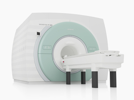
From Siemens Medical Systems;
The MAGNETOM 7T is designed as an open research platform. 7T MRI provides anatomical detail at the submillimiter scale, enhanced contrast mechanisms, outstanding spectroscopy performance, ultra-high resolution functional imaging (fMRI), multinuclear whole-body MRI and functional information.
This ultra high field (UHF) MRI device is a research system and not cleared, approved or licensed in any jurisdiction for patient examinations.
Device Information and Specification
CLINICAL APPLICATION
Whole body
CONFIGURATION
Compact
7 Tesla
High-performance, ultra high field coils available. Integration and support for coil developments.
CHANNELS (min. / max. configuration)
32, optional 8 channels TX array
Chemical shift imaging, single voxel spectroscopy, multinuclear imaging optional
IMAGING TECHNIQUES
iPAT, mSENSE and GRAPPA (image, k-space), noncontrast angiography, plaque imaging, radial motion compensation
FOV
40 x 40 x 30 cm³ less than 8% nonlinearity
BORE DIAMETER
or W x H
60 cm
TABLE CAPACITY
200 kg
MAGNET WEIGHT (gantry included)
35017 kg
DIMENSION H*W*D (gantry included)
320 x 240 x 317,5 cm
5-GAUSS FRINGE FIELD
7.9 m / 5.6 m
CRYOGEN USE
Zero boil off rate
COOLING SYSTEM
Water
up to 200 T/m/s
MAX. AMPLITUDE
up to 70 mT/m
Up to 3rd order shim coils, user configurable B0 shim ? B0 maps and ROI definition
POWER REQUIREMENTS
2000 Volts, 650A
spacer
 
Further Reading:
  Basics:
MAGNETOM 7T Product Brochure
   by www.healthcare.siemens.com    
  News & More:
Ultra-high-field MRI may allow earlier diagnosis of Parkinson's disease
Wednesday, 5 March 2014   by www.sciencedaily.com    
Feasibility of Using Ultra-High Field (7 T) MRI for Clinical Surgical Targeting
Thursday, 17 May 2012   by www.plosone.org    
Ultrahigh-Field MRI May Detect Additional Pathology in EAE
Sunday, 20 October 2013   by www.msdiscovery.org    
MRI Resources 
Health - Calculation - Pediatric and Fetal MRI - Hospitals - MRCP - Breast MRI
 
previous      21 - 25 (of 41)     next
Result Pages : [1]  [2 3 4 5 6 7 8 9]
 Random Page
 
Share This Page
FacebookTwitterLinkedIn

MR-TIP    
Community   
User
Pass
Forgot your UserID/Password ?    


MRI is trending to low field magnets :
reduced costs will lead to this change 
AI will close the gap to high field 
only in remote areas 
is only temporary 
never 

Look
      Ups


MR-TIP.com uses cookies! By browsing MR-TIP.com, you agree to our use of cookies.

Magnetic Resonance - Technology Information Portal
Member of SoftWays' Medical Imaging Group - MR-TIP • Radiology-TIP • Medical-Ultrasound-Imaging • 
Copyright © 2003 - 2024 SoftWays. All rights reserved. [ 20 April 2024]
Terms of Use | Privacy Policy | Advertising
 [last update: 2024-02-26 03:41:00]