| Info
Sheets |
| | | | | | | | | | | | | | | | | | | | | | | | |
 | Out-
side |
| | | | |
|
| | | | |
Result : Searchterm 'Tesla' found in 3 terms [ ] and 38 definitions [ ] and 38 definitions [ ] ]
| | 1 - 5 (of 41) nextResult Pages : [1] [2 3 4 5 6 7 8 9] |  | |  | Searchterm 'Tesla' was also found in the following services: | | | | |
| |  |
| Tesla |  |
| |
|
(T) The SI unit of magnetic flux density.
Definition: 1 T is the field intensity generating 1 newton of force per ampere of current per meter of conductor.
The tesla unit value is defined as a field strength of 1 weber per square meter of area, where 1 weber represents 1 x 10⁸ (100 000 000) flux lines.
One T is equal to 10 000 gauss, the older (CGS) unit.
A field of 1 tesla is quite strong, the Earth's magnetic flux density, at its surface, is about 50 micro teslas (μT). The slew rate of MRI devices is measured in mT/m/msec or T/m/sec. | | | | | | • Share the entry 'Tesla':    | | | | | | | | | |  Further Reading: Further Reading: | | Basics:
|
|
News & More:
| |
| |
| | | Searchterm 'Tesla' was also found in the following services: | | | | |
| | |
| |
|
(Mn-DPDP) This agent, mangafodipir trisodium, is a hepatocyte specific MRI contrast agent. Manganese is very toxic, so it has to be chelated and put in the form of a vitamin B6 analog, which is taken up by normal hepatocytes to some extent.
Teslascan® was developed in the early 1980's, went through clinical trials in the early 1990's, and was approved in 1997. One problem with assessing the efficacy of this agent is the fact that the phase III trials finished in the early 1990's, and the techniques used for MR today are very different from the techniques used almost a decade ago.
This contrast agent shortens the T1 relaxation time. On T1 weighted pictures it makes a normal liver look brighter. Since metastases, for example, do not generally take up this agent, the contrast between the enhancing liver and the non-enhancing lesions will increase on T1 weighted pictures. It does not have much effect on T2 weighted images. Drug Information and Specification T1, Predominantly positive enhancement PHARMACOKINETIC Hepatobiliary, pancreatic, adrenal DOSAGE 5 µmol/kg, 0.5 ml/kg PREPARATION Finished product DEVELOPMENT STAGE Approved PRESENTATION Vials of 100 ml DO NOT RELY ON THE INFORMATION PROVIDED HERE, THEY ARE
NOT A SUBSTITUTE FOR THE ACCOMPANYING PACKAGE INSERT! Distribution Information TERRITORY TRADE NAME DEVELOPMENT
STAGE DISTRIBUTOR | | | |
• View the DATABASE results for 'Teslascan®' (4).
| | | | | | Further Reading: | Basics:
|
|
News & More:
| |
| |
| | | | | |
| |
|
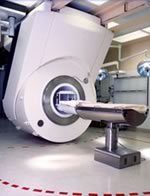
Device Information and Specification
CLINICAL APPLICATION
Whole body
CONFIGURATION
Mobile compact
Whole body, intra-operative head, neck volume, atlas head//neck vascular quadrature phased array, spine quadrature, C/T/L spine phased array, small joint, large joint, TMJ bilateral, shoulder phased array, extremity quadrature volume, wrist, hand quadrature, general purpose flexible, pelvis/abdomen phased array, body quadrature, phased array flexible, breast bilateral
IMAGING MODES
Localizer, single slice, multislice, volume
| | | |
• View the DATABASE results for 'iMotion™ 1.5 Tesla Magnet' (2).
| | | | |
| | | Searchterm 'Tesla' was also found in the following services: | | | | |
| |  |
| |
|
The subacute risks and side effects of magnetic and RF fields (for patients and staff) have been intensively examined for a long time, but there have been no long-term studies following persons who have been exposed to the static magnetic fields used in MRI. However, no permanent hazardous effects of a static magnetic field exposure upon human beings have yet been demonstrated.
Temporary possible side effects of high magnetic and RF fields:
•
Varying magnetic fields can induce so-called magnetic phosphenes that occur when an individual is subject to rapid changes of 2-5 T/s, which can produce a flashing sensation in the eyes. This temporary side effect does not seem to damage the eyes. Static field strengths used for clinical MRI examinations vary between 0.2 and 3.0 tesla;; field changes during the MRI scan vary in the dimension of mT/s. Experimental imaging units can use higher field strengths of up to 14.0 T, which are not approved for human use.
•
The Radio frequency pulses mainly produce heat, which is absorbed by the body tissue. If the power of the RF radiation is very high, the patient may be heated too much. To avoid this heating, the limit of RF exposure in MRI is up to the maximum specific absorption rate (SAR) of 4 W/kg whole body weight (can be different from country to country). For MRI safety reasons, the MRI machine starts no sequence, if the SAR limit is exceeded.
•
Very high static magnetic fields are needed to reduce the conductivity of nerves perceptibly. Augmentation of T waves is observed at fields used in standard imaging but this side effect in MRI is completely reversible upon removal from the magnet. Cardiac arrhythmia threshold is typically set to 7-10 tesla. The magnetohydrodynamic effect, which results from a voltage occurring across a vessel in a magnetic field and percolated by a saline solution such as blood, is irrelevant at the field strengths used.
The results of some animal and cellular studies suggest the possibility that electromagnetic fields may act as co-carcinogens or tumor promoters, but the data are inconclusive.
Up to 45 tesla, no important effects on enzyme systems have been observed. Neither changes in enzyme kinetics, nor orientation changes in macromolecules have been conclusively demonstrated.
There are some publications associating an increase in the incidence of leukemia with the location of buildings close to high-current power lines with extremely low-frequency (ELF) electromagnetic radiation of 50-60 Hz, and industrial exposure to electric and magnetic fields but a transposition of such effects to MRI or MRS seems unlikely.
Under consideration of the MRI safety guidelines, real dangers or risks of an exposure with common MRI field strengths up to 3 tesla as well as the RF exposure during the MRI scan, are not to be expected.
For more MRI safety information see also Nerve Conductivity,
Contraindications, Pregnancy
and Specific Absorption Rate.
See also the related poll result: ' In 2010 your scanner will probably work with a field strength of' | | | |
• View the DATABASE results for 'MRI Risks' (9).
| | |
• View the NEWS results for 'MRI Risks' (3).
| | | | | | Further Reading: | Basics:
|
|
News & More:
| |
| |
| | | Searchterm 'Tesla' was also found in the following services: | | | | |
| | |
| |
|
| | | |
• View the DATABASE results for 'Mangafodipir Trisodium' (6).
| | |
• View the NEWS results for 'Mangafodipir Trisodium' (1).
| | | | | | Further Reading: | Basics:
|
|
News & More:
| |
| |
| | | | |
| | | |
|
| |
 | Look
Ups |
| |