Magnetic Resonance - Technology Information Portal Welcome to MRI Technology
Info
  Sheets

Out-
      side


 'T1' 
SEARCH FOR    
 
  2 3 5 A B C D E F G H I J K L M N O P Q R S T U V W X Y Z
Result : Searchterm 'T1' found in 10 terms [] and 165 definitions []
previous     31 - 35 (of 175)     next
Result Pages : [1 2]  [3 4 5 6 7 8 9 10 11 12 13 14 15 16 17 18 19 20 ... ]
Searchterm 'T1' was also found in the following services: 
spacer
News  (18)  Resources  (5)  Forum  (54)  
 
Fast Spin EchoForum -
related threadsInfoSheet: - Sequences - 
Intro, 
Overview, 
Types of, 
etc.
 
Fast Spin Echo Diagram 
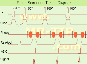
(FSE) In the pulse sequence timing diagram, a fast spin echo sequence with an echo train length of 3 is illustrated. This sequence is characterized by a series of rapidly applied 180° rephasing pulses and multiple echoes, changing the phase encoding gradient for each echo.
The echo time TE may vary from echo to echo in the echo train. The echoes in the center of the K-space (in the case of linear k-space acquisition) mainly produce the type of image contrast, whereas the periphery of K-space determines the spatial resolution. For example, in the middle of K-space the late echoes of T2 weighted images are encoded. T1 or PD contrast is produced from the early echoes.
The benefit of this technique is that the scan duration with, e.g. a turbo spin echo turbo factor / echo train length of 9, is one ninth of the time. In T1 weighted and proton density weighted sequences, there is a limit to how large the ETL can be (e.g. a usual ETL for T1 weighted images is between 3 and 7). The use of large echo train lengths with short TE results in blurring and loss of contrast. For this reason, T2 weighted imaging profits most from this technique.
In T2 weighted FSE images, both water and fat are hyperintense. This is because the succession of 180° RF pulses reduces the spin spin interactions in fat and increases its T2 decay time. Fast spin echo (FSE) sequences have replaced conventional T2 weighted spin echo sequences for most clinical applications. Fast spin echo allows reduced acquisition times and enables T2 weighted breath hold imaging, e.g. for applications in the upper abdomen.
In case of the acquisition of 2 echoes this type of a sequence is named double fast spin echo / dual echo sequence, the first echo is usually density and the second echo is T2 weighted image. Fast spin echo images are more T2 weighted, which makes it difficult to obtain true proton density weighted images. For dual echo imaging with density weighting, the TR should be kept between 2000 - 2400 msec with a short ETL (e.g., 4).
Other terms for this technique are:
Turbo Spin Echo
Rapid Imaging Spin Echo,
Rapid Spin Echo,
Rapid Acquisition Spin Echo,
Rapid Acquisition with Refocused Echoes
 
Images, Movies, Sliders:
 Lumbar Spine T2 FSE Sagittal  Open this link in a new window
    

Courtesy of  Robert R. Edelman
 MRI - Anatomic Imaging of the Foot  Open this link in a new window
    
SlidersSliders Overview

 Lumbar Spine T2 FSE Axial  Open this link in a new window
    

Courtesy of  Robert R. Edelman
 
spacer
 
• Related Searches:
    • Lumbar Spine MRI
    • Image Contrast Characteristics
    • Pulse Sequence Timing Diagram
    • Liver Imaging
    • Turbo Spin Echo
 
Further Reading:
  Basics:
MYELIN-SELECTIVE MRI: PULSE SEQUENCE DESIGN AND OPTIMIZATION
   by www.imaging.robarts.ca    
Advances in Magnetic Resonance Neuroimaging
Friday, 27 February 2009   by www.ncbi.nlm.nih.gov    
  News & More:
New MR sequence helps radiologists more accurately evaluate abnormalities of the uterus and ovaries
Thursday, 23 April 2009   by www.eurekalert.org    
Spin echoes, CPMG and T2 relaxation - Introductory NMR & MRI from Magritek
2013   by www.azom.com    
MRI Resources 
Safety pool - Spine MRI - Artifacts - Movies - MRI Reimbursement - Breast MRI
 
Longitudinal Relaxation Time
 
The T1 time constant, which determines the rate at which excited protons return to equilibrium within the lattice. The longitudinal relaxation time is a measure of the time taken for spinning protons to realign with the external magnetic field. The magnetization will grow after excitation from zero to a value of about 63% of its final value in a time of T1.

See also T1 Time.
 
Images, Movies, Sliders:
 Brain MRI Images T1  Open this link in a new window
 Sagittal Knee MRI Images T1 Weighted  Open this link in a new window
 
spacer

• View the DATABASE results for 'Longitudinal Relaxation Time' (5).Open this link in a new window

MRI Resources 
Sequences - Intraoperative MRI - Process Analysis - General - MR Myelography - Cardiovascular Imaging
 
LumenHance®InfoSheet: - Contrast Agents - 
Intro, Overview, 
Characteristics, 
Types of, 
etc.MRI Resource Directory:
 - Contrast Agents -
 
Oral administration of this liposomal encapsulated manganese chloride results in a reduction of the T1 relaxation time causing enhanced signal on T1 weighted MR images. Bracco received marketing approval in December 1997 in the United States, but marketing state is still uncertain.

See also Classifications, Characteristics, etc.
Drug Information and Specification
NAME OF COMPOUND
MnCl2, Manganese chloride tetrahydrate
CENTRAL MOIETY
Mn2+
CONTRAST EFFECT
T1, Predominantly positive enhancement
Paramagnetic
PHARMACOKINETIC
Gastrointestinal
PREPARATION
Powder for reconstitution
INDICATION
Bowel marking
DEVELOPMENT STAGE
For sale
DISTRIBUTOR
See below
DO NOT RELY ON THE INFORMATION PROVIDED HERE, THEY ARE
NOT A SUBSTITUTE FOR THE ACCOMPANYING PACKAGE INSERT!
Distribution Information
TERRITORY
TRADE NAME
DEVELOPMENT
STAGE
DISTRIBUTOR
LumenHance®
for sale
spacer

• View the DATABASE results for 'LumenHance®' (4).Open this link in a new window

Searchterm 'T1' was also found in the following services: 
spacer
News  (18)  Resources  (5)  Forum  (54)  
 
Magnevist®InfoSheet: - Contrast Agents - 
Intro, Overview, 
Characteristics, 
Types of, 
etc.MRI Resource Directory:
 - Contrast Agents -
 
Magnevist® is a paramagnetic ionic contrast agent for use in magnetic resonance imaging. Contrast enhanced MRI with Magnevist® allows additional diagnostic information of tumors, inflammation and vascular lesions and the determination or differentiation of such lesions.
The contrast enhancing effect is produced by the di-N-methylglucamine salt of gadopentetate (Gd-DTPA), the gadolinium complex of diethylenetriamine pentaacetic acid. Magnevist® has the strongest effect on T1 weighted images, by increasing T1 signal intensity in tissues where Magnevist® has accumulated.

WARNING: NEPHROGENIC SYSTEMIC FIBROSIS Gadolinium-based contrast agents increase the risk for nephrogenic systemic fibrosis (NSF) in patients with acute or chronic severe renal insufficiency (glomerular filtration rate less than 30 mL/min/1.73m2), or acute renal insufficiency of any severity due to the hepato-renal syndrome or in the perioperative liver transplantation period.

See also Ionic Intravenous Contrast Agents and Gadopentetate Dimeglumine.
Drug Information and Specification
CENTRAL MOIETY
Gd3+
CONTRAST EFFECT
T1, Predominantly positive enhancement
r1=3.4, r2=3.8, B0=1.0T
PHARMACOKINETIC
Intravascular, extracellular, renal excretion
1960 mosm/kgH2O
CONCENTRATION
0.5 mol/L
DOSAGE
0.1-0.3 mmol/kg / 0.2-0.6 mL/kg
PREPARATION
Finished product
INDICATION
Neuro/whole body
DEVELOPMENT STAGE
For sale
DISTRIBUTOR
See below
PRESENTATION
Vials of 5, 10, 15, 20 and 100 mL bulk package
Pre-filled syringes of 10, 15 and 20 mL
DO NOT RELY ON THE INFORMATION PROVIDED HERE, THEY ARE
NOT A SUBSTITUTE FOR THE ACCOMPANYING PACKAGE INSERT!
Distribution Information
TERRITORY
TRADE NAME
DEVELOPMENT
STAGE
DISTRIBUTOR
USA, Canada
Magnevist®
for sale
Asia
Magnevist®
for sale
EU
Magnevist®
suspended
Turkey
Magnevist®, Magnograf
for sale
Australia
Magnevist®
for sale
spacer

• View the DATABASE results for 'Magnevist®' (7).Open this link in a new window

 
Further Reading:
  Basics:
Magnevist Prescription Drug Discount Sources
   by www.institutedc.org    
Important Drug Warning for Gadolinium-Based Contrast Agents
Wednesday, 12 September 2007   by www.ismrm.org    
MAGNEVIST ® (brand of gadopentetate dimeglumine) Injection - WARNING: NEPHROGENIC SYSTEMIC FIBROSIS
May 2009   by berlex.bayerhealthcare.com    
Magnevist Package Insert
2000
  News & More:
EMA's final opinion confirms restrictions on use of linear gadolinium agents in body scans
Friday, 21 July 2017   by www.ema.europa.eu    
Gadolinium-containing contrast agents: removal of Omniscan and iv Magnevist, restrictions to the use of other linear agents
Friday, 5 January 2018   by www.gov.uk    
FDA Drug Safety Communication: FDA warns that gadolinium-based contrast agents (GBCAs) are retained in the body; requires new class warnings
Tuesday, 19 December 2017   by www.fda.gov    
Spurious Hypocalcemia After Omniscan- or OptiMARK-Enhanced Magnetic Resonance Imaging: An Algorithm for Minimizing a False-Positive Laboratory Value
October 2004   by www.findarticles.com    
MRI Resources 
Sequences - Blood Flow Imaging - Education - Portals - MRI Accidents - Contrast Enhanced MRI
 
O-SCAN™InfoSheet: - Devices -
Intro, 
Types of Magnets, 
Overview, 
etc.
 
www.fonar.com/standup.htm www.fonar.com/standup.htm O-scan is manufactured and distributed by Esaote SpA
O-scan is a compact, dedicated extremity MRI system designed for easy installation and high throughput. The complete system fits in a 9' x 10' room, doesn't need for RF or magnetic shielding and it plugs in the wall. The 0.31T permanent magnet along with dual phased array RF coils, and advanced imaging protocols provide outstanding image quality and fast 25 minute complete examinations.
Esaote North America is the exclusive distributor of the O-scan system in the USA.
Device Information and Specification
CLINICAL APPLICATION
Dedicated Extremity
CONFIGURATION
Closed
Dual phased array knee, hand, foot//ankle/elbow
PULSE SEQUENCES
SE, HSE, HFE, GE, 2dGE, ME, IR, STIR, Stir T2, GESTIR, TSE, TME, FSE STIR, FSE (T1, T2), X-Bone, Turbo 3DT1, 3D SHARC, 3D SST1, 3D SST2
IMAGING MODES
2D, 3D multi-plane, half echo, half scan, real time
TR
10 - 10,000 msec.
TE
6 - 220 msec.
SINGLE SLICE
0.1 sec.
MULTI SLICE
0.1 sec.
14 cm
2D: 2mm - 10 mm, 3D: 0.6 - 10 mm
MEASURING MATRIX
512 x 512 max.
PIXEL INTENSITY
4,096 grey levels
MAGNET TYPE
Permanent - NdFeB
MAGNET WEIGHT
2,733 lbs
POWER REQUIREMENTS
100/110/200/220/230/240
STRENGTH
20 mT/m
5 GAUSS FRINGE FIELD, radial/axial
67 cm / 75 cm
passive
spacer
MRI Resources 
Musculoskeletal and Joint MRI - MRI Training Courses - Cochlear Implant - Nerve Stimulator - MR Myelography - Patient Information
 
previous      31 - 35 (of 175)     next
Result Pages : [1 2]  [3 4 5 6 7 8 9 10 11 12 13 14 15 16 17 18 19 20 ... ]
 Random Page
 
Share This Page
FacebookTwitterLinkedIn

MR-TIP    
Community   
User
Pass
Forgot your UserID/Password ?    


MRI is trending to low field magnets :
reduced costs will lead to this change 
AI will close the gap to high field 
only in remote areas 
is only temporary 
never 

Look
      Ups


MR-TIP.com uses cookies! By browsing MR-TIP.com, you agree to our use of cookies.

Magnetic Resonance - Technology Information Portal
Member of SoftWays' Medical Imaging Group - MR-TIP • Radiology-TIP • Medical-Ultrasound-Imaging • 
Copyright © 2003 - 2024 SoftWays. All rights reserved. [ 25 April 2024]
Terms of Use | Privacy Policy | Advertising
 [last update: 2024-02-26 03:41:00]