 | Info
Sheets |
| | | | | | | | | | | | | | | | | | | | | | | | |
 | Out-
side |
| | | | |
|
| | | | | |  | Searchterm 'Spatial Resolution' was also found in the following services: | | | | |
|  |  |
| |
|
Device Information and Specification
CLINICAL APPLICATION
Whole body
CONFIGURATION
Cylindrical Wide Short Bore
SE, FE, IR, FastSE, FastIR, FastFLAIR, Fast STIR, FastFE, FASE, Hybrid EPI, Multi Shot EPI; Angiography: 2D(gate/non-gate)/3D TOF, SORS-STC
IMAGING MODES
Single, multislice, volume study
TE
8 msec min. SE; 0.9 msec min. FE
less than 0.011 (256x256)
1.0 min. 2-DFT: 0.2 min. 3-DFT
32-1024, phase;; 64-1024, freq.
65.5 cm, patient aperture
4050 kg (bare magnet incl. L-He)
POWER REQUIREMENTS
380/400/415/440/480 V
COOLING SYSTEM TYPE
Closed-loop water-cooled
Liquid helium: approx. less than 0.05 L/hr
Passive, active, auto-active
| | | | | |  Further Reading: Further Reading: | News & More:
|
|
| |
| | | Searchterm 'Spatial Resolution' was also found in the following service: | | | | |
| | |
| |
|
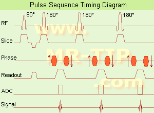
(FSE) In the pulse sequence timing diagram, a fast spin echo sequence with an echo train length of 3 is illustrated.
This sequence is characterized by a series of rapidly applied 180° rephasing pulses and multiple echoes, changing the phase encoding gradient for each echo.
The echo time TE may vary from echo to echo in the echo train. The echoes in the center of the K-space (in the case of linear k-space acquisition) mainly produce the type of image contrast, whereas the periphery of K-space determines the spatial resolution. For example, in the middle of K-space the late echoes of T2 weighted images are encoded. T1 or PD contrast is produced from the early echoes.
The benefit of this technique is that the scan duration with, e.g. a turbo spin echo turbo factor / echo train length of 9, is one ninth of the time. In T1 weighted and proton density weighted sequences, there is a limit to how large the ETL can be (e.g. a usual ETL for T1 weighted images is between 3 and 7). The use of large echo train lengths with short TE results in blurring and loss of contrast. For this reason, T2 weighted imaging profits most from this technique.
In T2 weighted FSE images, both water and fat are hyperintense. This is because the succession of 180° RF pulses reduces the spin spin interactions in fat and increases its T2 decay time. Fast spin echo (FSE) sequences have replaced conventional T2 weighted spin echo sequences for most clinical applications. Fast spin echo allows reduced acquisition times and enables T2 weighted breath hold imaging, e.g. for applications in the upper abdomen.
In case of the acquisition of 2 echoes this type of a sequence is named double fast spin echo / dual echo sequence, the first echo is usually density and the second echo is T2 weighted image. Fast spin echo images are more T2 weighted, which makes it difficult to obtain true proton density weighted images. For dual echo imaging with density weighting, the TR should be kept between 2000 - 2400 msec with a short ETL (e.g., 4).
Other terms for this technique are:
Turbo Spin Echo
Rapid Imaging Spin Echo,
Rapid Spin Echo,
Rapid Acquisition Spin Echo,
Rapid Acquisition with Refocused Echoes
| | | | | |
• View the DATABASE results for 'Fast Spin Echo' (31).
| | | | | | Further Reading: | | Basics:
|
|
News & More:
| |
| |
| | | | | |
| |
|
(FWHM) A commonly used measure of the width at half the maximum value of peaked functions such as spectral lines or slice profiles and important measure of the quality of an imaging device and its spatial resolution.
As the name states, the FWHM is measured by identifying the points on the signal curve, which are half the maximum value. The horizontal distance between these two points is called the FWHM. For a spectral line, this will be proportional to 1/T2. | | | |
• View the DATABASE results for 'Full Width at Half Maximum' (3).
| | | | | | Further Reading: | | Basics:
|
|
News & More:
| |
| |
| | | Searchterm 'Spatial Resolution' was also found in the following services: | | | | |
| | |
| |
|
The use of MR spectroscopy for acquiring functional activation of the brain. There are two possible approaches:
In the first, localized spectra of brain water are acquired and subtle changes in these spectra reflect the biophysical water environment. Changes in T2 due to deoxyhaemoglobin concentration may be detected in this way.
The disadvantages of poor spatial resolution are to some extent offset by the high signal to noise ratio SNR of the spectroscopic data.
An alternative approach is to use MR spectroscopy directly to detect metabolites that are altered by brain activation. These include lactate and glucose. Such experiments have inherently poor spatial and temporal resolution, but do give a direct indication of the metabolic response of the brain to functional activation. | | | | | |
| | | Searchterm 'Spatial Resolution' was also found in the following service: | | | | |
| | |
| |
|
(fMRI) Functional magnetic resonance imaging is a technique used to determine the dynamic brain function, often based on echo planar imaging, but can also be performed by using contrast agents and observing their first pass effects through brain tissue. Functional magnetic resonance imaging allows insights in a dysfunctional brain as well as into the basic workings of the brain.
The in functional brain MRI most frequently used effect to assess brain function is the blood oxygenation level dependent contrast ( BOLD) effect, in which differential changes in brain perfusion and their resultant effect on the regional distribution of oxy- to deoxyhaemoglobin are observable because of the different 'intrinsic contrast media' effects of the two haemoglobin forms. Increased brain activity causes an increased demand for oxygen, and the vascular system actually overcompensates for this, increasing the amount of oxygenated haemoglobin. Because deoxygenated haemoglobin attenuates the MR signal, the vascular response leads to a signal increase that is related to the neural activity.
Functional imaging relates body function or thought to specific locations where the neural activity is taking place. The brain is scanned at low resolution but at a fast rate (typically once every 2-3 seconds). Structural MRI together with fMRI provides an anatomical baseline and best spatial resolution.
Interactions can also be seen from the motor cortex to the cerebellum or basal ganglia in the case of a movement disorder such as ataxia. For example: by a finger movement the briefly increase in the blood circulation of the appropriate part of the brain controlling that movement, can be measured. | | | |
• View the DATABASE results for 'Functional Magnetic Resonance Imaging' (8).
| | |
• View the NEWS results for 'Functional Magnetic Resonance Imaging' (15).
| | | | | | Further Reading: | Basics:
|
|
News & More:
| |
| |
| | | | |
| | | |
|
| |
 | Look
Ups |
| |


