Magnetic Resonance - Technology Information Portal Welcome to MRI Technology
Info
  Sheets

Out-
      side


 'Signal to Noise Ratio' 
SEARCH FOR    
 
  2 3 5 A B C D E F G H I J K L M N O P Q R S T U V W X Y Z
Result : Searchterm 'Signal to Noise Ratio' found in 1 term [] and 49 definitions []
previous     21 - 25 (of 50)     next
Result Pages : [1]  [2 3 4 5 6 7 8 9 10]
Searchterm 'Signal to Noise Ratio' was also found in the following services: 
spacer
News  (3)  
 
Image Resolution
 
The image resolution is the level of detail of an image and a measurement of image quality. Higher resolution means more image detail, for example when two structures 1 mm apart are distinguishable in an image, this picture has a higher resolution than an image where they are not to distinguish.
More data points in an MR image (with same FOV) will decrease the pixel size, but not accurately improve the resolution because the different MRI sequences influence the contrast and the discernment of different tissues. With high contrast and optimal signal to noise ratio, the image resolution is depend on FOV and number of data points of a picture, but T2* effects have an additional influence.
spacer

• View the NEWS results for 'Image Resolution' (1).Open this link in a new window.
 
Further Reading:
  Basics:
The Basics of MRI
   by www.cis.rit.edu    
  News & More:
Optimizing Musculoskeletal MR
   by rad.usuhs.mil    
Searchterm 'Signal to Noise Ratio' was also found in the following services: 
spacer
Radiology  (2) Open this link in a new windowUltrasound  (8) Open this link in a new window
Imaging CoilInfoSheet: - Coils - 
Intro, 
Overview, 
etc.MRI Resource Directory:
 - Coils -
 
Imaging coils are radio frequency coils used in magnetic resonance imaging for sending and/or receiving electromagnetic radiation. Several MR imaging coils in different shapes and sizes are necessary for different body parts and to handle individual applications.
For Example:
Birdcage coils provide high homogeneity and good signal to noise ratio (SNR) in brain MRI scans.
Surface coils with small coil diameter and higher SNR enable to image the temporomandibular joints (TMJ).
The implemented body coil allows the scanning of body parts with large field of views.
Micro coils are available to image finger joints.
High performance phased array coils are today state-of-the-art for a wide range of applications from head, spine, knee or shoulder to cardiac MRI.
For different types of coils see Volume Coil, Sense Coil, Array Coil, Surface Coil, and Bird Cage Coil.

See also the related poll result: '3rd party coils are better than the original manufacturer coils'
spacer

• View the DATABASE results for 'Imaging Coil' (7).Open this link in a new window


• View the NEWS results for 'Imaging Coil' (9).Open this link in a new window.
 
Further Reading:
  Basics:
Radio-frequency Coil Selection for MR Imaging of the Brain and Skull Base1
   by radiology.rsnajnls.org    
MRI Resources 
Knee MRI - Resources - IR - Shoulder MRI - MRI Centers - Supplies
 
Low Field MRIForum -
related threads
 
Advantages of low field imaging are the small-sized 5 Gauss fringe field and therefore the less static magnetic field exposure for the surrounding area, as well as less contraindications causing lower risks for the MRI safety by implemented metal and magnetic devices and equipment.
Low field systems are sometimes for restricted use, e.g. dedicated extremity scanner or open MRI devices. Open MRI devices equipped with permanent magnets are well-suited for MR guided interventions because these machines combine the lower magnetic fields of this type of magnets and the better patient access of open MRI scanner.
In some cases, the contrast of different tissues is better at lower field strength, depending on their T1 or T2 relaxation times. The disadvantage of the lower signal to noise ratio are a poor resolution and a longer scan time for a good image quality.

See also Claustrophobia, Contraindications and MRI Safety.

See also the related poll result: 'In 2010 your scanner will probably work with a field strength of'
spacer

• View the DATABASE results for 'Low Field MRI' (8).Open this link in a new window


• View the NEWS results for 'Low Field MRI' (5).Open this link in a new window.
 
Further Reading:
  Basics:
Improving portable low-field MRI image quality through image-to-image translation using paired low- and high-field images
Friday, 1 December 2023   by www.nature.com    
  News & More:
Safety of Bedside Portable Low-Field Brain MRI in ECMO Patients Supported on Intra-Aortic Balloon Pump
Friday, 18 November 2022   by www.mdpi.com    
Researchers at the University of Tsukuba develop a portable MRI system specifically for identifying wrist cartilage damage among athletes, providing a convenient means of early detection and treatment of injuries
Tuesday, 26 April 2022   by www.tsukuba.ac.jp    
This bizarre looking helmet can create better brain scans
Friday, 11 February 2022   by www.sciencedaily.com    
A low-cost and shielding-free ultra-low-field brain MRI scanner
Tuesday, 14 December 2021   by www.nature.com    
Portable MRI provides life-saving information to doctors treating strokes
Thursday, 5 August 2021   by news.yale.edu    
Synaptive Evry, an MRI for Any Space, Cleared by FDA
Thursday, 30 April 2020   by www.medgadget.com    
World's First Portable MRI Cleared by FDA
Monday, 17 February 2020   by www.medgadget.com    
Introducing a point-of-care MRI system
Tuesday, 29 October 2019   by healthcare-in-europe.com    
Opportunities in Interventional and Diagnostic Imaging by Using High-performance Low-Field-Strength MRI
Tuesday, 1 October 2019   by pubs.rsna.org    
Portable 'battlefield MRI' comes out of the lab
Thursday, 30 April 2015   by physicsworld.com    
Portable MRI could aid wounded soldiers and children in the third world
Thursday, 23 April 2015   by phys.org    
Searchterm 'Signal to Noise Ratio' was also found in the following services: 
spacer
News  (3)  
 
Lung ImagingMRI Resource Directory:
 - Lung Imaging -
 
Lung imaging is furthermore a challenge in MRI because of the predominance of air within the lungs and associated susceptibility issues as well as low signal to noise of the inflated lung parenchyma. Cardiac and respiratory triggered or breath hold sequences allow diagnostic imaging, however a comparable image quality with computed tomography is still difficult to achieve.
Assumptions for lung MRI:
•
Low signal to noise ratio of the inherently low lung proton density.
•
Cardiac and respiratory motion artifacts.
•
Magnetic susceptibility effects of large magnetic field gradients.
•
Very short transverse relaxation times and significant diffusion yielding short T2 (30-70 msec), short T2* (1-3 msec), and additional long T1 relaxation times (1300-1500 msec).
•
The extreme short T2 values are responsible for a fast signal decay during a single shot readout, resulting in blurring.

The current trends in MRI are the use of new imaging technologies and increasingly powerful magnetic fields. Among these technologies are parallel imaging techniques as well as ventilation agents like hyperpolarized helium for the use as an inert inhalational contrast agent to study lung ventilation properties. With hyperpolarized gases clear images of the lungs can be obtained without using a large magnetic field (see also back projection imaging). Single shot sequences (e.g. TSE or Half Fourier Acquisition Single Shot Turbo Spin Echo HASTE) used in lung MR imaging benefits from parallel imaging techniques due to reduced relaxation time effects during the echo train and therefore reduced image blurring as well as reduced motion artifacts.
In the future, more effective contrast agents may provide an alternative solution to the need for high field MRI. Dynamic contrast enhanced MRI perfusion has demonstrated a potential in the diagnosis of pulmonary embolism or to characterize lung cancer and mediastinal tumors. 3D contrast enhanced magnetic resonance angiography of the thoracic vessel.

See also the related poll result: 'MRI will have replaced 50% of x-ray exams by'
 
Images, Movies, Sliders:
 Anatomic Imaging of the Lungs  Open this link in a new window
      

Courtesy of  Robert R. Edelman
 Normal Lung Gd Perfusion MRI  Open this link in a new window
      

Courtesy of  Robert R. Edelman

 MRI Thorax Basal Plane  Open this link in a new window
 
Radiology-tip.comradLung Scintigraphy
spacer

• View the DATABASE results for 'Lung Imaging' (7).Open this link in a new window


• View the NEWS results for 'Lung Imaging' (3).Open this link in a new window.
 
Further Reading:
  Basics:
A safer approach for diagnostic medical imaging
Monday, 29 September 2014   by www.eurekalert.org    
Parallel Lung Imaging(.pdf)
  News & More:
Chest MRI a viable alternative to chest CT in COVID-19 pneumonia follow-up
Monday, 21 September 2020   by www.healthimaging.com    
CT Imaging Features of 2019 Novel Corona virus (2019-nCoV)
Tuesday, 4 February 2020   by pubs.rsna.org    
Polarean Imaging Phase III Trial Results Point to Potential Improvements in Lung Imaging
Wednesday, 29 January 2020   by www.diagnosticimaging.com    
Low Power MRI Helps Image Lungs, Brings Costs Down
Thursday, 10 October 2019   by www.medgadget.com    
Chest MRI Using Multivane-XD, a Novel T2-Weighted Free Breathing MR Sequence
Thursday, 11 July 2019   by www.sciencedirect.co    
Researchers Review Importance of Non-Invasive Imaging in Diagnosis and Management of PAH
Wednesday, 11 March 2015   by lungdiseasenews.com    
New MRI Approach Reveals Bronchiectasis' Key Features Within the Lung
Thursday, 13 November 2014   by lungdiseasenews.com    
MRI techniques improve pulmonary embolism detection
Monday, 19 March 2012   by medicalxpress.com    
  News & More:
Partnership with VIDA to streamline adoption of advanced MRI of the lungs
Monday, 11 September 2023   by www.itnonline.com    
Searchterm 'Signal to Noise Ratio' was also found in the following services: 
spacer
Radiology  (2) Open this link in a new windowUltrasound  (8) Open this link in a new window
MAGNETOM Skyra
 
www.healthcare.siemens.com/magnetic-resonance-imaging/0-35-to-1-5t-mri-scanner/magnetom-skyra/ 
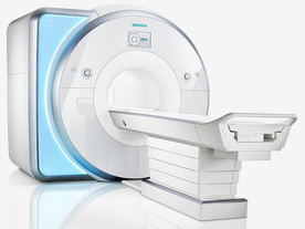
From Siemens Medical Systems; Received FDA clearance in 2010.
MAGNETOM Skyra is a top-of-the-line, patient friendly wide bore 3 Tesla MRI system.
The system is equipped with the Tim 4G and Dot system (Total imaging matrix and Day optimizing throughput), to enhance both productivity and image quality with the complete range of advanced applications for clinical routine and research. Tim 4G features lighter, trimmer MRI coils that take up less space inside the magnet but deliver a high coil element density with increased signal to noise ratio and the possibility to use high iPAT factors.
Device Information and Specification
CLINICAL APPLICATION
Whole Body
CONFIGURATION
Open bore
3 Tesla
Head, spine, torso/ body coil, neurovascular, cardiac, neck, shoulder, knee, wrist, foot//ankle and multi-purpose flex coils. Peripheral vascular, breast, shoulder.
CHANNELS (min. / max. configuration)
48, 64, 128
Chemical shift imaging, single voxel spectroscopy
IMAGING TECHNIQUES
iPAT, mSENSE and GRAPPA (image, k-space),CAIPIRINHA (k-space), noncontrast angiography, plaque imaging, radial motion compensation, Dixon
MINIMUM TR
3D T1 spoiled GRE: 0.95 (256 matrix)
MINIMUM TE
3D T1 spoiled GRE: 0.22 (256 matrix), Ultra-short TE
FOV
0.5 - 50 cm
BORE DIAMETER
or W x H
At isocenter: L-R 70 cm, A-P (with table) 55 cm
TABLE CAPACITY
250 kg
MAGNET WEIGHT (gantry included)
5768 kg
DIMENSION H*W*D (gantry included)
173 x 231 x 219 cm
5-GAUSS FRINGE FIELD
2.6 m / 4.6 m
CRYOGEN USE
Zero boil off rate, approx. 10 years
COOLING SYSTEM
Water; single cryogen, 2 stage refrigeration
up to 200 T/m/s
MAX. AMPLITUDE
45 mT/m
3 linear with 20 coils, 5 nonlinear 2nd-order
POWER REQUIREMENTS
380 / 400 / 420 / 440 / 460 / 480 V, 3-phase + ground; 110 kVA
spacer
MRI Resources 
Jobs pool - MRI Reimbursement - Mobile MRI - MR Guided Interventions - Sequences - Chemistry
 
previous      21 - 25 (of 50)     next
Result Pages : [1]  [2 3 4 5 6 7 8 9 10]
 Random Page
 
Share This Page
FacebookTwitterLinkedIn

MR-TIP    
Community   
User
Pass
Forgot your UserID/Password ?    


New acceleration techniques will :
reduce scan times 
cause artifacts 
increase expenses 
be useful if you have a lot of experience 
doesn't do much 
never heard of 

Look
      Ups


MR-TIP.com uses cookies! By browsing MR-TIP.com, you agree to our use of cookies.

Magnetic Resonance - Technology Information Portal
Member of SoftWays' Medical Imaging Group - MR-TIP • Radiology-TIP • Medical-Ultrasound-Imaging • 
Copyright © 2003 - 2024 SoftWays. All rights reserved. [ 23 April 2024]
Terms of Use | Privacy Policy | Advertising
 [last update: 2024-02-26 03:41:00]