 | Info
Sheets |
| | | | | | | | | | | | | | | | | | | | | | | | |
 | Out-
side |
| | | | |
|
| | | | |
Result : Searchterm 'Shot' found in 8 terms [ ] and 28 definitions [ ] and 28 definitions [ ] ]
| | previous 11 - 15 (of 36) nextResult Pages : [1 2] [3 4 5 6 7 8] |  | |  | Searchterm 'Shot' was also found in the following services: | | | | |
| |  |
| |
|
Ultrasound imaging is the primary fetal monitoring modality during pregnancy, nevertheless fetal MRI is increasingly used to image anatomical regions and structures difficult to see with sonography. Given its long record of safety, utility, and cost-effectiveness, ultrasound will remain the modality of first choice in fetal screening. However, MRI is beginning to fill a niche in situations where ultrasound does not provide enough information to diagnose abnormalities before the baby's birth. Magnetic resonance imaging of the fetus provides multiplanar views also in sub-optimal positions, better characterization of anatomic details of e.g. the fetal brain, and information for planning the mode of delivery and airway management at birth.
Indications:
•
Examinations of the placenta
Modern fetal MRI requires no sedatives or muscle relaxants to control fetal movement. Ultrafast MRI techniques (e.g., single shot techniques like Half Fourier Acquisition Single shot Turbo spin Echo HASTE) enable images to be acquired in less than one second to eliminate fetal motion. Such technology has led to increased usage of fetal MRI, which can lead to earlier diagnosis of conditions affecting the baby and has proven useful in planning fetal surgery and designing postnatal treatments. As MR technology continues to improve, more advances in the prenatal diagnosis and treatment of fetal abnormalities are to expect. More advances in in-utero interventions are likely as well. Eventually, fetal MRI may replace even some prenatal tests that require invasive procedures such as amniocentesis.
For Ultrasound Imaging (USI) see Fetal Ultrasound at Medical-Ultrasound-Imaging.com. | | | | | | | | | | | | |  Further Reading: Further Reading: | | Basics:
|
|
News & More:
|  |
Advances in medical imaging enable visualization of white matter tracts in fetuses
Wednesday, 12 May 2021 by www.eurekalert.or | | |
Fetal CMR Detects Congenital Heart Defects, Changes Treatment Decisions
Monday, 29 March 2021 by www.diagnosticimaging.com | | |
MRI scans more precisely define and detect some abnormalities in unborn babies
Friday, 12 March 2021 by www.eurekalert.org | | |
Ultrasound and Magnetic Resonance Imaging of Agenesis of the Corpus Callosum in Fetuses: Frontal Horns and Cavum Septi Pellucidi Are Clues to Earlier Diagnosis
Monday, 29 June 2020 by pubmed.ncbi.nlm.nih.gov | | |
MRI helps predict preterm birth
Tuesday, 15 March 2016 by www.eurekalert.org | | |
3-T MRI advancing on ultrasound for imaging fetal abnormalities
Monday, 20 April 2015 by www.eurekalert.org | | |
Babies benefit from pioneering 'miniature' MRI scanner in Sheffield
Friday, 24 January 2014 by www.telegraph.co.uk | | |
Ultrasensitive Detector Pinpoints Big Problem in Tiny Fetal Heart
Tuesday, 6 April 2010 by www.sciencedaily.com | | |
Real-time MRI helps doctors assess beating heart in fetus
Thursday, 29 September 2005 by www.eurekalert.org |
|
| |
| | | Searchterm 'Shot' was also found in the following services: | | | | |
| | |
| |
|
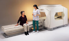
From Toshiba America Medical Systems Inc.;
the Ultra™ system was developed to help healthcare providers be more competitive by delivering greater patient comfort and a broad range of clinical capabilities, says Anita Bowler, product manager, MRI Business Unit, Toshiba America Medical Systems Inc With its unique, powerful gradient technology, the Ultra™ performs advanced clinical studies and consistently provides high-resolution images that are typically associated with high field MRI systems. At the same time, the Ultra™ offers a truly open feeling that makes patients more relaxed, especially those with claustrophobic tendencies.
Device Information and Specification CLINICAL APPLICATION Whole Body Quadrature, solenoid and multi-channel configurations SE, FE, IR, FastSE, FastIR, FastFLAIR, Fast STIR, FastFE, FASE, Hybrid EPI, Multi Shot EPI, Single shot EPI diffusion, True SSFP, SuperFASE; Angiography: 2D(gate/non-gate)/3D TOF, SORS-STC, Black Blood MRA POWER REQUIREMENTS 380/400/415/440/480 V COOLING SYSTEM TYPE Cryogenless | | | | | |
| | | | | |
| |
|
Quick Overview
Please note that there are different common names for this artifact.
HELP
Fast scan techniques
Patient movement during the scans are often an imaging problem. Artifacts from patient movement are widely varied due to a dependence when during k-space filling the motion occurs. When the patient moving causes only in the last few seconds of the scan at that time the outside edges of K-space were being filled, and as a result the artifact does not overly affect the image (there are only fine lines).

Image Guidance
| | | | | | | Further Reading: | News & More:
|
|
| |
| | | Searchterm 'Shot' was also found in the following services: | | | | |
| | |
| |
|

'Next generation MRI system 1.5T CHORUS developed by ISOL Technology is optimized for both clinical diagnostic imaging and for research development.
CHORUS offers the complete range of feature oriented advanced imaging techniques- for both clinical routine and research. The compact short bore magnet, the patient friendly design and the gradient technology make the innovation to new degree of perfection in magnetic resonance.'
Device Information and Specification
CLINICAL APPLICATION
Whole body
Spin Echo, Gradient Echo, Fast Spin Echo,
Inversion Recovery ( STIR, Fluid Attenuated Inversion Recovery), FLASH, FISP, PSIF, Turbo Flash ( MPRAGE ),TOF MR Angiography, Standard echo planar imaging package (SE-EPI, GE-EPI), Optional:
Advanced P.A. Imaging Package (up to 4 ch.), Advanced echo planar imaging package,
Single Shot and Diffusion Weighted EPI, IR/FLAIR EPI
STRENGTH
20 mT/m (Upto 27 mT/m)
| | | |
• View the DATABASE results for 'CHORUS 1.5T™' (2).
| | | | |
| | | Searchterm 'Shot' was also found in the following services: | | | | |
| | |
| |
|

(EPI) Echo planar imaging is one of the early magnetic resonance imaging sequences (also known as Intascan), used in applications like diffusion, perfusion, and functional magnetic resonance imaging. Other sequences acquire one k-space line at each phase encoding step. When the echo planar imaging acquisition strategy is used, the complete image is formed from a single data sample (all k-space lines are measured in one repetition time) of a gradient echo or spin echo sequence (see single shot technique) with an acquisition time of about 20 to 100 ms.
The pulse sequence timing diagram illustrates an echo planar imaging sequence from spin echo type with eight echo train pulses. (See also Pulse Sequence Timing Diagram, for a description of the components.)
In case of a gradient echo based EPI sequence the initial part is very similar to a standard gradient echo sequence. By periodically fast reversing the readout or frequency encoding gradient, a train of echoes is generated.
EPI requires higher performance from the MRI scanner like much larger gradient amplitudes. The scan time is dependent on the spatial resolution required, the strength of the applied gradient fields and the time the machine needs to ramp the gradients.
In EPI, there is water fat shift in the phase encoding direction due to phase accumulations. To minimize water fat shift (WFS) in the phase direction fat suppression and a wide bandwidth (BW) are selected. On a typical EPI sequence, there is virtually no time at all for the flat top of the gradient waveform. The problem is solved by "ramp sampling" through most of the rise and fall time to improve image resolution.
The benefits of the fast imaging time are not without cost. EPI is relatively demanding on the scanner hardware, in particular on gradient strengths, gradient switching times, and receiver bandwidth. In addition, EPI is extremely sensitive to image artifacts and distortions. | | | |
• View the DATABASE results for 'Echo Planar Imaging' (19).
| | |
• View the NEWS results for 'Echo Planar Imaging' (1).
| | | | | | Further Reading: | Basics:
|
|
| |
| | | | |
| | | |
|
| |
 | Look
Ups |
| |


