 | Info
Sheets |
| | | | | | | | | | | | | | | | | | | | | | | | |
 | Out-
side |
| | | | |
|
| | | | |
Result : Searchterm 'Shielding' found in 8 terms [ ] and 18 definitions [ ] and 18 definitions [ ] ]
| | previous 21 - 25 (of 26) nextResult Pages : [1 2] [3 4 5 6] |  | |  | Searchterm 'Shielding' was also found in the following services: | | | | |
| |  |
| |
|

It is important to remember when working around a superconducting magnet that the magnetic field is always on. Under usual working conditions the field is never turned off. Attention must be paid to keep all ferromagnetic items at an adequate distance from the magnet. Ferromagnetic objects which came accidentally under the influence of these strong magnets can injure or kill individuals in or nearby the magnet, or can seriously damage every hardware, the magnet itself, the cooling system, etc..
See MRI resources Accidents.
The doors leading to a magnet room should be closed at all times except when entering or exiting the room. Every person working in or entering the magnet room or adjacent rooms with a magnetic field has to be instructed about the dangers. This should include the patient, intensive-care staff, and maintenance-, service- and cleaning personnel, etc..
The 5 Gauss limit defines the 'safe' level of static magnetic field exposure. The value of the absorbed dose is fixed by the authorities to avoid heating of the patient's tissue and is defined by the specific absorption rate.
Leads or wires that are used in the magnet bore during imaging procedures, should not form large-radius wire loops. Leg-to-leg and leg-to-arm skin contact should be prevented in order to avoid the risk of burning due to the generation of high current loops if the legs or arms are allowed to touch. The patient's skin should not be in contact with the inner bore of the magnet.
The outflow from cryogens like liquid helium is improbable during normal operation and not a real danger for patients.
The safety of MRI contrast agents is tested in drug trials and they have a high compatibility with very few side effects. The variations of the side effects and possible contraindications are similar to X-ray contrast medium, but very rare. In general, an adverse reaction increases with the quantity of the MRI contrast medium and also with the osmolarity of the compound.
See also 5 Gauss Fringe Field, 5 Gauss Line, Cardiac Risks, Cardiac Stent, dB/dt, Legal Requirements, Low Field MRI, Magnetohydrodynamic Effect, MR Compatibility, MR Guided Interventions, Claustrophobia, MRI Risks and Shielding. | | | | | | | | | • For this and other aspects of MRI safety see our InfoSheet about MRI Safety. | | | • Patient-related information is collected in our MRI Patient Information.
| | | | | | | | | |  Further Reading: Further Reading: | | Basics:
|
|
News & More:
| |
| |
| | | Searchterm 'Shielding' was also found in the following service: | | | | |
| | |
| |
|
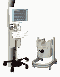
From MagneVu;
The MagneVu 1000 is a compact, robust, and portable, permanent magnet MRI system and operates without special shielding or costly site preparation.
This MRI device utilizes a patented non-homogeneous magnetic field image acquisition method to achieve high performance imaging. The MagneVu 1000 MRI scanner is designed for MRI of the extremities with the current specialty areas in diabetes and rheumatoid arthritis. Easy access is afforded for claustrophobic, pediatric, or limited mobility patients. In August 1998
FDA marketing clearance and other regulatory approvals have been received. Until 2008, over 130 devices in the US are in use. Some further developments of MagneVu's extremity scanner are: 'truly Plug n' Play MRI™' and iSiS ( which adds wireless capability to the second generation MV1000-XL).
Device Information and Specification IMAGING MODES 3-dimensional multi-echo data acquisition | | | |
• View the DATABASE results for 'MagneVu 1000' (3).
| | | | | | Further Reading: | News & More:
|
|
| |
| |  | MRI Safety Resources | | | | |
| | | |
| |
|
Quick Overview
NAME
Moire fringes, moire
DESCRIPTION
Superimposed signals of different phases
A moiré pattern is an interference pattern created for example when two grids are overlaid at an angle, or when they have slightly different mesh sizes. The human visual system creates an imaginary pattern of roughly horizontal dark and light bands, the moiré pattern that appears to be superimposed on the lines.
In MRI, the appearance of moiré fringes can be caused by a variety of reasons e.g., inhomogeneity of the main magnetic field caused by a defect shielding ( interference with RF pulses), interferences produced by aliasing, and interferences of echoes from different excitation modes (with different echo times).

Image Guidance
| | | | | | | Further Reading: | Basics:
|
|
| |
| | | Searchterm 'Shielding' was also found in the following services: | | | | |
| | |
| |
|


O-scan is manufactured and distributed by Esaote SpA
O-scan is a compact, dedicated extremity MRI system designed for easy installation and high throughput. The complete system fits in a 9' x 10' room, doesn't need for RF or magnetic shielding and it plugs in the wall. The 0.31T permanent magnet along with dual phased array RF coils, and advanced imaging protocols provide outstanding image quality and fast 25 minute complete examinations.
Esaote North America is the exclusive distributor of the O-scan system in the USA.
Device Information and Specification CLINICAL APPLICATION Dedicated Extremity
PULSE SEQUENCES
SE, HSE, HFE, GE, 2dGE, ME, IR, STIR, Stir T2, GESTIR, TSE, TME, FSE STIR, FSE ( T1, T2), X-Bone, Turbo 3DT1, 3D SHARC, 3D SST1, 3D SST2 2D: 2mm - 10 mm, 3D: 0.6 - 10 mm POWER REQUIREMENTS 100/110/200/220/230/240 | | | | | |
| | | Searchterm 'Shielding' was also found in the following service: | | | | |
| | |
| |
|
| | | |
• View the DATABASE results for 'Shielded Gradient Coils' (4).
| | | | | | Further Reading: | Basics:
|
|
| |
| | | | |
| | | |
|
| |
 | Look
Ups |
| |


